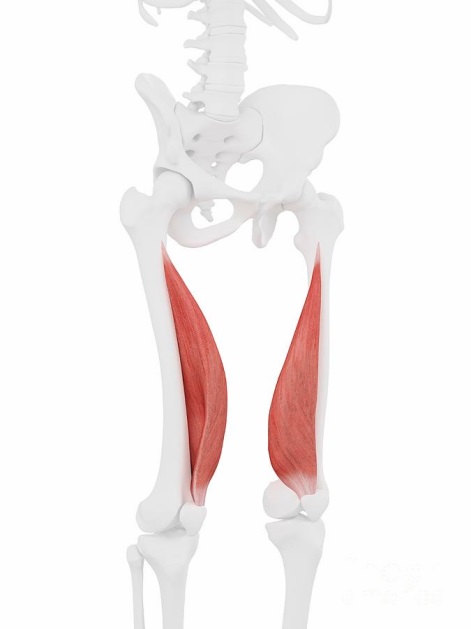
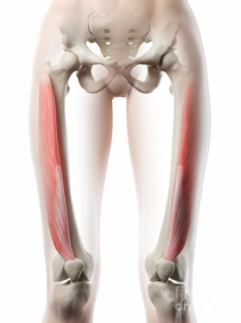
The quadriceps muscle (m. quadriceps) is a muscle that largely determines the appearance of the lower extremity. The quadriceps play a critical role in uprightness and, as one of the leading “anti-gravity” muscles, keeps the knee from bending under the whole body’s weight. The function of the rectus femoris muscle is to turn the hip at the hip joint and extend the leg at the knee joint. It helps to keep the head of the femur inside the acetabulum. The rectus femoris muscle occupies the medial position and is the primary motor.
The vastus medialis muscle is classified as a secondary motor and performs a similar function. The function of the vastus medialis muscle is to participate in extending the knee along with the other muscles that make up the quadriceps muscle. It also contributes to proper patella gliding and maintains the mobility of the entire knee area. The function of the vastus lateralis muscle is the extensor of the tibia at the knee joint. It is attached to the greater trochanter, providing elasticity for the movements of the rectus muscle. The function of the vastus intermedius muscle is also to extend the tibia at the knee joint; it runs in line with the tibial tuberosity.
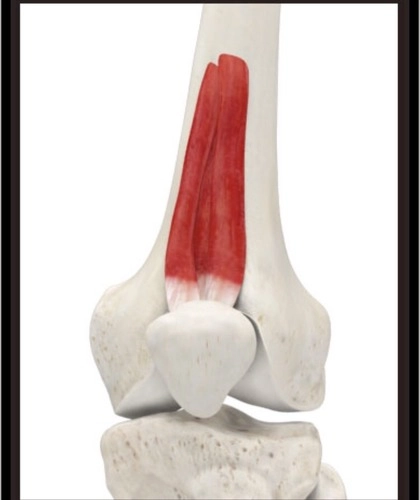
The articular knee muscle is a small muscle of the anterior thigh group, a flat plate consisting of several well-defined muscle bundles. It belongs to the tertiary motors and has the function of elevating the patellar sac and preventing deformation of the synovial sheath. In addition, the joint muscle provides preservation of the knee joint by mediating a collective interaction with the quadriceps femoris muscle.
The sartorius muscle is the longest in the human body. This muscle bends the leg at the hip and knee joints: it rotates the tibia inward and the thigh outward. Doing so takes part in throwing the leg behind the leg. It participates in straightening the hip and prevents it from turning inward during squats. It also engages in medial rotation of the knee joint.
- rectus femoris muscle – secondary mover, antagonist;
- articularis genus muscle – quaternary mover, stabilizer;
- vastus medialis muscle – tertiary mover, synergists;
- vastus lateralis muscle – tertiary mover, synergists;
- vastus intermedius muscle – tertiary mover, synergists;
- musculus sartorius – primary mover, agonist.
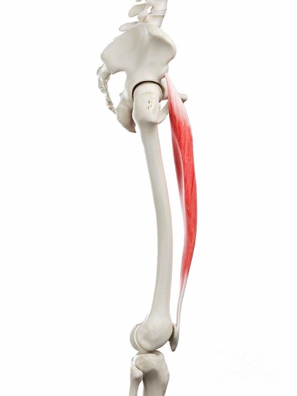
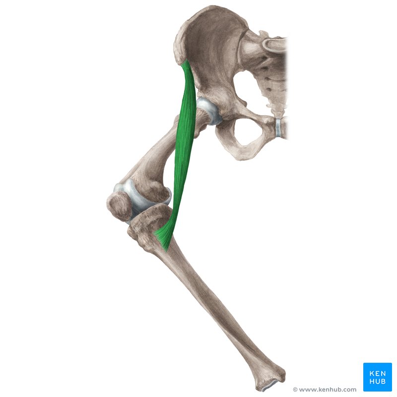
Pes anserinus (goosefoot) refers to the tendons of three thigh muscles: the tailor muscle, the thin muscle, and the semitendinosus muscle. The three goosefoot tendons are superficial to the knee’s medial collateral ligament (MCL). The portacarpal and lean muscles are the adductors of the hip (that is, they drive the hip to the medial axis of the body). The semitendinosus muscle is part of the hamstrings group and is located on the posterior surface of the thigh. Together, these three muscles are the flexors and pronators of the knee.
The anserine pouch, or goosefoot pouch, is a fluid-filled bubble. It secretes synovial fluid to reduce friction between tissues and serves as a shock absorber for bones, ligaments, and muscles. Inflammation of the pouch does not occur suddenly but develops over a long period. Traumatization of pes anserinus leads to such consequences as strain when walking, inability to run and walk for long distances, and impaired biomechanics of the lower extremities. If treatment is delayed, the infection can spread to other areas and limit the use of the entire limb. In addition, knee injury is always one of the highest pain indexes, resulting in impaired gait and limb flexion.
References
Lee JH, Kim KJ, Jeong YG, et al. Pes anserinus and anserine bursa: anatomical study. Anatomy & Cell Biology. 2014;47(2):127-131. Web.
Netter F. Atlas of Human Anatomy (7th ed.). Saunders, 2019.
Sanami K. The Femoral Nerve. Teach Me Anatomy. Web.
Vascovic J. Quadriceps femoris muscle. Kenhub. Web.