Write-up for the onion root tip experiment
The Cell Cycle and Mitosis
Growth of an organism requires increase in cell mass, duplication of genetic material and a division. Eukaryotic cells contain highly organized genetic material contained in the nucleus, a membrane bound organelle. The fundamental unit of genetic information is chromatin, composed of double stranded DNA, making complex with histone proteins in highly wounded ribbon like shapes appearing as “beads on a sting” or nucleosomes. The DNA is a large molecule consisting of helical units of phosphates and deoxyribose sugar to which nitrogenous bases, adenine (A), thiamine (T), cytosine (C) and guanine (G) are linked. The sequence of the bases determines the nature of protein it codes. DNA in chromatin is also inheritable unit and the process of mitosis ensures that exact copy of DNA in chromatin distributes between the daughter cells. A cell requiring 24 hours to complete the division cycle, undergoes non-dividing stages of G1 (Gap 1)-S (Synthesis)-G2 (Gap 2) lasting 23 hours during which cell mass increases and DNA of every chromosome replicates, and a mitotic dividing stage (M) of 1 hour, in which sister chromatids of each chromosome separates, one each going to the daughter cells. Mitosis is comprised of five sequential phases culminating in cytokinesis, means changes in cytoplasm that include division of cell proper. During interphase that lies between the two cell divisions encompassing G1-S-G2 stages, the chromatin material is not clearly visible under microscope. A distinguishable feature is a dark spot in the nucleus called nucleolus. Microtubules originate from a pair of centrioles at the onset of prophase. During prophase chromatin starts to condense to chromosomes and nuclear membrane disappears. Each chromosome contains two chromatids held together by centromere. During prometaphase microtubules together with some specialized proteins anchor the centromeres and make a complex called kinetochore microtubules, and chromosomes begin to move. The metaphase is characteristic of an alignment of chromosomes in a line in the middle of the cells called equator. Kinetochore microtubules condense to form spindle fibers holding the centromeres together. Another region of microtubule formation away from centromere starts from the poles. The paired chromatids separate at the point of centromere during anaphase and move to the opposite directions approaching poles with the help of spindle fibers and polar microtubules. During telophase, chromatids already clustered in the poles, uncoil and new nuclear membrane is formed. Chromatids reorganize to the chromatin network. The exact number and quantity of parental cell chromosomes distribute between the two daughter cells.
Onion Root Tip Investigation
Problem Question
In dividing cells of an onion root tip, what % of the cell cycle is spent in interphase and what % is spent in mitosis (all stages combined)? It is hypothesized that a large proportion of the cells would undergo mitotic nuclear division, and relative to interphase or non-dividing cells more number of cells would be seen in one of the four phases – prophase, metaphase, anaphase and telophase. The reason for making this hypothesis is that the roots continue to profusely grow both apically and laterally in search of nutrients and water and root tips are mainly responsible for absorption, hence the cells should be continuously rejuvenating by repeated cycles of cell division.
Procedure
Temporary acetocarmine stained mounts of onion root tip can be prepared by (a) remove old roots from onion bulb and put the bulb on a bottle full of water and within 3-4 days new roots will appear, (b) when 2-3 cm long cut new roots from the tips preferably early in the morning, (c) keep the roots for 24 hours in fixative-preservative, Formal-Alcohol fixative solution containing formalin, ethyl alcohol and glacial acetic acid, (d) transfer them to 1:1 solution of conc. HCl and 95% alcohol for 10 minutes, (e) keep them for 10 min in fixative-dehydrating, Carnoy’s fluid comprising absolute alcohol, chloroform and glacial acetic acid, and (d) transfer the root tips in 70% alcohol until used. A tip is then kept on a clean slide and a drop of acetocarmine stain is added onto it. By warming the slide on flame the excess stain can be evaporated. Repeat straining and evaporating the specimen for a couple of cycles. The tip can be squashed using forceps and cover it with coverslip. Gently press the coverslip with blotting paper to make uniform spreading of squash and to remove excess stain. Finally, examine first at low power and selected field can be magnified under high power using a compound microscope. From among several such slides, the best one showing various stages of mitosis and with optimal nuclear staining has been picked up for enumeration of the total number of stained cells, and cells that are either not dividing i.e. at interphase (prominent dark spot of nucleoli, defined nuclear membrane and no organized chromosome) or dividing i.e. at one of the stages of mitosis (anucleolated cells, loss of nuclear membrane, organized chromosomal structures moving at different positions, spindle fibers etc).

Results
Analysis
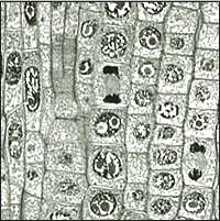
The results from this photograph indicate that over half the cells were at interphase and were not dividing, whereas from the remaining dividing cells most were at prophase or in transition from interphase to prophase. There was none showing metaphase or telophase and only a minor proportion of the cells exhibited being in anaphase. Hence a total 47% dividing cells were present in the optical field and 53% cells were non-dividing at interphase.
Conclusion
The hypothesis that cells were profusely mitotically dividing was not supported, rather it appears that the cells were more in the resting non-dividing stage. The fact that visibly some cells could not be distinguished from interphase to prophase and some cells did not stain properly, our assumption on % cells at interphase may slightly change. But this confusion has no implication on the % of dividing cells at later stages, like about 7% cells were present at anaphase stage. Telophase stage is also difficult to distinguish from prophase unless the cell plate can be clearly demarcated in the slide. Taken together, having only minor proportion of the cells in anaphase and none in metaphase and telophase is suggestive of poor mitosis in the given root tip cells. It is possible that in other root tip squashes there might be more dividing cells than resting cells but to make a statement as to whether the given hypothesis is accepted or rejected, more root tips need to be investigated and the data should be statistically treated.
Meiosis
In meiosis chromosomes of diploid cells (2n) divide in four haploid (n) daughter cells. Meiosis is responsible for gamete formation and it also generates genetic variations through recombination. Meiosis takes place in two steps – meiosis I and II. In meiosis I, prophase I is a long stage in which chromatin network shows condensation to chromosome and nucleoli tend to disappear, but different from mitotic-prophase, two homologous chromosomes (one maternal and one paternal) lie side-by-side and this pair of chromosomes is known as bivalents. Each bivalent splits longitudinally into two sister chromatids and hence bivalents become tetrads. At this level genetic recombination takes place at chiasmata. Later, chromosomes condense further and partially separate. In metaphase I, bivalents lay side-by-side in equatorial plane and two members of the pair face opposite poles. The kinetochore microtubules anchor each chromosome rather than chromatid. Each chromosome with two chromatids separate and move to opposite poles by spindle fibers during anaphase I. At telophase I chromosome loose their identity and daughter nuclei is formed and nucleoli reappear. In meiosis II, prophase II chromosomes reappear as two chromatids while nucleolus and nuclear membrane begin to disappear. The chromatids lie in equatorial plane in metaphase II and each chromatid gets attached to spindle fibers and pulled towards the opposite poles. Telophase II observes chromatids (now chromosomes) to reorganize into chromatin network again, reappearance of nucleolus and formation of haploid nucleus. Thereafter, cytokinesis takes place to produce gametes.
Control experiment
The above root tip experiment is not a controlled investigation but it is possible to include a control experiment to evaluate the variable component of the experiment. In controls, the mitosis should be arrested such that the chromatin replication during S phase is not affected as the later condition would be lethal. Among many chemicals colchicines are known to restrict nucleus division at metaphase. They do so because of interference with microtubule synthesis and consequently not allowing the equatorial arranged chromosomes of metaphase to segregate and move towards poles. In the given experiment onion bulbs can be treated with 0.05% colchicine in water before new root tips are cut for fixation. Due to arrest of mitosis at metaphase cells would not divide and also look identical in photomicrographs. Any variation in mitosis due to various factors can be controlled using this experiment.
Five basic differences between mitosis and meiosis
- One cell produce two daughter cells in mitosis whereas four daughter cells in meiosis.
- Meiosis is reduction division which means the diploid cells (2n) divide into haploid cells (n), whereas in mitosis the progeny cells remain diploid.
- The genetic recombination takes place at chiasmata from crossing over of the paired chromosomes in prophase I in meiosis, whereas the genetic identity is maintained and no exchange of DNA material takes place in case of mitosis.
- Mitosis is accomplished in only one step whereas meiosis takes two independent steps called meiosis I and II.
- The homologous chromosomes one each acquired from each parent make pair in metaphase I in meiosis, but in mitosis (metaphase) all the chromosomes do not pair but make a line regardless of whether homologous or not.
Discussion
Introduction to Biology lecture course is a good extension of mitosis and meiosis assignments and is not repetition of earlier assignments. In the assignment over 50% of the root tip cells were non-dividing and only 7% cells proceeded up to mitosis anaphase. More root tip analysis would be necessary to make a better judgment.