Vision
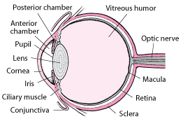
The eye made up of the eyeball, ciliary muscles, and nerve cells is the organ for vision. The sclera has a thin mucous layer that protects the cornea. Light enters the eye through the cornea and passes through the iris to the retina where it is focused. According to Starr, the iris controls the amount of light entering the eye by either contracting or enlarging. This is aided by the papillary and dilator muscles. The iris focuses light to the retina (Starr, 435).

The retina contains the macula which has photoreceptor cells that provide nourishment alongside the blood vessels (Beverly, 342). Photoreceptor cells are linked to the optic nerve fibers and make the image more visual. They also change the observed images into impulses that are the transmitted to the brain through the optic fibers. Photoreceptor cells are classified as either rods or cones. The cones found in the macula are responsible for the central vision of the image including its color. Rods which are numerous and more sensitive to light help in vision during darkness (Cecie, 213).
Works Cited
Starr, Cecie, & Beverly McMillan. ”Human Biology”. 9th edition.USA: Brooks Cole. 2012. Print.