Introduction
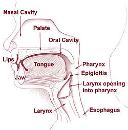
The esophagus (or gullet) is a 10 inches long midline muscular tube lying at the front of the spine, and it links the throat to the stomach. Furthermore, the esophagus is positioned before the right side of the spine after the windpipe in the upper layer of the chest, and behind the heart in the lower part of the chest (Kantsevoy 9).
The esophagus goes down through three compartments: the neck, the chest, and the abdomen. This chain of movement has led to the standard anatomical splitting up of cervical, thoracic, and abdominal subdivisions. The esophagus, both proximally and distally, is alleviated by bony, cartilaginous, or membranous formations (Braden, and Daniela 2006).

Structure of the Esophagus
The esophagus connects with the pharynx, and begins its formation at the cricoids cartilage opposite cervical vertebra (Dofka 61). It goes down into the chest at the stage of the sterna notch and moves inside the chest cavity on the fore plane of the posterior mediastinum. It moves down the spine and stops at the esophagogastric junction between the thoracic inlet and the diaphragm.
Location of the Esophagus
The esophagus is located slightly to the right of the midline, especially in its course through the thorax, where it descends along the right side of the aorta.
Minor variations of Esophagus
Three minor variations are present in the esophagus. The first variation is present at the bottom of the neck. The second variation is present at the thoracic vertebra. The third variation takes place when the esophagogastric junction (cardia) is positioned lateral to the xiphoid process of the sternum.
Divisions of the Esophagus
The esophagus is approximately 23-27 cm long in diameter, and is divided into three parts:
- Cervical part: it is positioned just at the anterior vertebral column in the neck.
- Thoracic part: this is the longest part of the esophagus and it can be found in the greater and subsequent mediastinum.
- Abdominal part: this is the shortest part of the esophagus and it is located in the peritoneal cavity. It lengthens to the cardiac orifice of the stomach from the diaphragm.
Constrictions of the esophagus
The esophagus has three normal anatomical constrictions, which are projected at the levels of specific vertebrae (Romer and Parsons 344). The constrictions are caused by adjacent structures that indent the esophagus by functional closure mechanisms. These constrictions are visible during gastroscopy, and the scope must be carefully maneuvered past them (normal width of the esophagus is approximately 20mm).
The three normal anatomical constrictions are:
- Upper Constriction: this corresponds to the esophageal inlet in the cervical part of the esophagus. It is located where the esophagus passes behind the cricoids cartilage and a maximum width of approximately 14mm.
- Middle constriction: this is located where the esophagus passes to the right of the aortic arch and thoracic aorta (at T4/T5). The maximum width is 14mm.
- Lower constriction: this is located at the start of the abdominal part of the esophagus, where it pierces the diaphragm (T10/T11). The abdominal part is normally occluded except during swallowing. The maximum width of the lower constriction is 14mm.
The esophagus also presents characteristics curves like an upper curve to the left (in the cervical part), a mid-level curve to the right (in the thoracic part, caused by the adjacent thoracic aorta), and a lower curve to the left (in the abdominal part). Additionally, the esophagus is slightly concave anteriorly in the sagittal plane, following the curvature of the vertebral column (thoracic kyphosis).
Works Cited
Braden, Kuo, and Daniela Urma. Esophagus – anatomy and development. GI Motility online, 2006. 10.1038. Print.
Dofka, Charline. Dental Terminology. New York, NY: Cengage Learning, 2000. Print.
Kantsevoy, Sergey. 2007 Johns Hopkins White Papers: Digestive Disorders. Baltimore, Maryland: Johns Hopkins Health, 2007. Print.
Romer, Sherwood, and Parsons Thomas. (2009). The Vertebrate Body. Philadelphia, PA: Holt-Saunders International. Print.