Introduction
In the advent of modern research facilities and advanced knowledge in the scientific field, many technological applications were developed as a means of improving people’s lives. Different advancements in scientific areas of discipline paved the way for beneficial inventions and innovations especially in the medical field. Radiology, for example, provided human beings the ability to explore his anatomy without surgery. There are many other examples of these advantageous technologies available to us in the modern times.
A very good example of technology’s benefits to human health is the development of a bone density test that only requires knowledge on radiation. It is important to check the density of the bone since it relates to a disease called osteoporosis. Osteoporosis is a disease of the bones characterized by the decrease in bone density due to lowering calcium levels and many other factors. It can be prevalent for a very long time, unnoticed by a patient suffering from it, unless a fracture transpires or unless detected by a bone density scan. This disease may be genetically caused or as an after-effect of medications such as steroids. It affects the way of life of the patient since it is directly linked to disability or death in the most serious cases (Hurd 2006).
The x-ray scan can immediately detect the occurrence of osteoporosis since the results on the x-ray film show significant differences from bones of a normal person in terms of diameter and other physical characteristics. Although x-ray can be used, a recent bone density scan was developed, called the dual energy x-ray absorptiometry scan (DEXA) that works similarly on the principle of radiation, but on a more precise level.
Bones
Anatomy
Bones, on the outside, are sturdy structures that protect and support the internal organs of the body and also affect the posture and overall shape of the body. It is composed of calcium and the inside of the bone is the literally the factory of blood cells. Ironically, the bone may seem to be lifeless but just like the skin and hair, it is continuously developing. This is evident in people who put extreme pressure to the bones such as weightlifters and athletes, who develop stronger bone structures. Women are more at risk in having osteoporosis since at the onset of menopause, there is a significant decrease in the levels of the hormone estrogen that prevents bone loss (“Osteoporosis”).
Bones develop constantly in length gradually from the inside. Ossification, or the process of bone growth, is characterized by the alteration of a weaker material of connective tissue by a sturdier bony tissue. Osteoblasts slowly develop first, forming an osteocyte. This continuous process soon creates a primary ossified center in the bone until it proceeds to the tip of the bones. As a result, there would be an increase in length followed by the development of a secondary ossified center in the epiphyses. Then, the weaker cartilaginous materials are substituted for by compact bony tissues. Development occurs early in the womb until adolescence when most of the time, the bones are considered to be mature. Growth in length ceases by this time as it is controlled by the growth hormone active during the puberty stage both in male and in females (“Bone and Development Growth”).
Breakdown of bones occurs naturally by the absorption of calcium in the bones. Vitamin D is a vitamin that enhances the absorption of calcium to the bones making it stronger. The National Osteoporosis Foundation and the National Institutes of Health both state the importance of Vitamin D in building stronger bones because of its very significant effect on calcium absorption (“Vitamin D’s Role”). Calcium does not only function in maintaining healthy bones but it is also needed by the body for transmitting electrical signals to and from the brain and also a component of lymph fluids. When the body is low in calcium, an immediate response is to take calcium stored in the bones. This leads to bone resorption and consequently, bone breakdown. As stated earlier, the menopausal stage in women and old age can also result to natural breakdown of the bones.
Osteoporosis Screening
In 2000, the statistics for older people suffering from osteoporosis rose at an alarming rate and the trend is increasing tremendously. Balbona (2000) reports that there are available screening tests for osteoporosis and interventions to prevent the onset of the disease. Experts believe that osteoporosis is a highly preventable disease that the medical industry has developed many suggestions for prevention. Patients’ levels of bone mineral densities (BMD) are constantly checked as this is a very good indicator of risk of osteoporosis occurrence. A value of BMD lower than “2.5 standard deviations from the mean BMD” is immediately termed as osteoporosis, whereas the BMD for normal bone density is within 1 standard deviation (Balbona 2000). “Osteoporosis diagnosis can be done by quantitative computed tomography (qCT), dual energy x-ray absorptiometry (DEXA) and single energy x-ray absorptiometry (SXA)” (Ott 2007). qCT makes use of comparison of measurements of the bone mineral density using t and z scores. Similarly, DEXA is quantitative in nature but is the most widely used to date as compared to SXA, which is an older method (Ott 2007).
Bone Densitometry
Technology
Kaufman (1999) states that bone density measurement started roughly during the 1940s making use of radiology, the most understood technology back then. The film that results from an x-ray scan did not do much good since at that time it was a very crude equipment. This led to the development of advanced bone densitometry devices that can clearly compare and isolate abnormalities of the bones compared to the normal. A Singh index was developed that takes advantage of the differences in the trabecular patterns of various bones. A grading system from 1-6 shows that fractures usually led to values of less than 3 when radiographed. Radiographic densitometry then commenced as another feat in the radiologic technology.
Dual Energy X-ray Absorptiometry (DEXA)
The DEXA makes use of low doses of radiation even lower than that used in chest x-rays. For instance, approximately 0.5-4.5 uSv is used by DEXA (DEXA RADIAITON SAFETY).The radioactive source of DEXA is an x-ray tube as compared to other scanning equipments that make use of gamma rays for instance. An x-ray generator is placed below while a detector or a device that can capture image is placed above. An invisible beam of x-rays having two different energy peaks are sent through the bone tissues. The two energy peaks are separately sent to the soft tissues and bones respectively. The values that can be obtained give the bone mineral density by subtracting the value of the energy peak of the soft tissue from the total. This is then sent to a computer for analysis and computation that later provides in display the results (“Bone Density Scan”).
Bone Mass Density
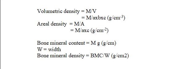
Densitometry works by the assumption that x-ray waves are absorbed by calcium ions, on the other hand, repelled by the bone tissues. The BMD is measured in grams/cm-3 of bone which makes up the volumetric density while a specific or given area of a bone is the areal density, measured in grams/cm-2 and usually done in the hip, wrist and the lower spine area. The mathematical equation to compute for the BMD is shown in Fig.1. DEXA can also be used to quantify the amount of fat and lean tissues of the whole body.

The densitometry machine takes a record of the computed results and compares it to a standard for osteoporosis since the main objective of the values is to correlate it to the risk of fracture. Values of the t-score and z-score are obtained as points of comparisons (Fig.2).

The t-scores reflect the “number of standard deviations (SD) above or below the young adult mean” (Kaufman 1999). This is in reference to a database previously input in the computer. Thus, the risk of fractures increases two-fold for every standard deviation that falls below the normal values. On the other hand, the z-scores suggest the difference between the values expected for the patient’s age bracket, also depending on the data previously in the database. The type of osteoporosis is also identified by the z-scores. Z values less than -1.5 are signs of secondary osteoporosis. Knowing these values easily benefit the patients suffering from osteoporosis since medications and preventions can be addressed as early as possible to avoid the disease to get worse (Kaufman 1999).
Radiation Safety
The Dubuque Internal Medicine in Iowa has released guidelines for adults who would like to undergo bone densitometry. Pregnant women are not allowed to take the test because the radiation may deliver negative effects to the developing baby. Also, patients who have had another x-ray or nuclear scan within the span of a week are not allowed. The patient should wear something comfortable like cotton shirts without any metals on it. Jeans and girdles shall be removed before taking the scan. Forty-eight hours before the exam, the patient is also not advised to take calcium, as this may interfere with the results that can be obtained leading to erroneous data. When everything is ready, the patient is asked to lie down on a flat area where an imaging device is positioned on top and the x-ray generator is placed below, depending on which part of the body is to be scanned. Scanning can take approximately 10 minutes per area and is painless like the typical chest x-ray. Radiologists and physicians are capable of analyzing the results and it may take some time before the report is relayed to the patient (Dubuque Internal Medicine).
Advantages and Disadvantages of DEXA
Exposure to some doses of radiation can be beneficial in terms of what is done in bone densitometry, but in large doses, radiation can cause damage to molecules inside the body, by formation of pyrimidine dimers in DNA, damaging membranes, inhibiting biological catabolic and anabolic pathways, inducing cancer, and eventually cause death This is the reason why the time and distance of exposure needs to be monitored both for the patient and the operator of the DEXA machine. Time of exposure to radiation must be kept short as possible to prevent absorption. Distance should also be kept and the use of Pb aprons is also recommended as shield (Maher 1997).
The DEXA is very cheap compared to other procedures that can be done, and again requires no surgery. Also, anaesthesia is no longer needed. Importantly, this machine is available in most health care facilities. What is very beneficial in this procedure is that no radiation is left in the patient’s body and this procedure is usually free from negative side effects. Yet, there is always the risk of triggering the formation of cancer cells. Unfortunately, the DEXA can only predict the risk of fracture instead of preventing it (“Bone Density Scan”).
Before the development of the dual energy x-ray absorptiometry, the single photon absorptiometry was made available first in the 1960s. This makes use of an isotope instead of an x-ray. Only a single beam of energy photons are made to pass through the tissues and the bone for quantification of the bone mineral density. Since it uses an isotope, the source has the tendency to decay, thus, constant replacement of the source is needed unlike in DEXA where the source is x-ray (Kaufman 1999). While DEXA is primarily used in scanning the lower spine and hips, the SEXA or single energy x-ray absorptiometry is widely used in the scanning of the peripheral areas of the body which include the wrist and heels. Since it only uses a single energy source, it is less accurate than the DEXA but it is beneficial in the peripheral regions of the body which do not necessarily need the power DEXA can provide.
Federal Legislation and Bone Densitometry
Bone Mass Measurement Act
July 1, 1998 marked the ratification of the Bone Mass Measurement Act that calls for the bone mass testing of Medicare beneficiaries all over the United States of America. This is a very efficient and beneficial move by the American health care system since the statistics for osteoporosis during those times were alarming enough that this kind of nationwide and extensive action is very much opportune (“Bone Mass Measurement”)..
Medicare beneficiaries capable of availing this service must be:
- “Any estrogen deficient woman who is determined by the physician or other nonphysician practitioner to be at clinical risk for osteoporosis based on her medical history or other findings. Even a woman on estrogen therapy can be considered to be estrogen deficient, especially if on an inadequate replacement dose or if noncompliant”. (Meckelnburg)
- “An individual with vertebral abnormalities as demonstrated on X-ray to be indicative of osteoporosis, low bone mass, or vertebral fracture”. (Meckelnburg)
- “An individual receiving or expected to receive glucocorticoid therapy equivalent to 7.5 mg prednisone or greater per day, for more than 3 months”. (Meckelnburg)
- “An individual with primary hyperparathyroidism” (Meckelnburg)
- “An individual being monitored to access the response to or efficacy of an FDA approved osteoporosis drug therapy” (Meckelnburg)
The Act did not only assist the beneficiaries on a one time basis, instead it was a continuous service goaled on long-term medication for them. Legislation also supported the Bone Mass Measurement Act by creating a string of policies. The Balanced Budget Act of 1997 details Medicare’s pledge to cover the procedures and techniques involved in bone mass measurement for the qualified beneficiaries. Title XVIII of the Social Security Act, on the other hand, protects the rights of Medicare as an organization and prevents abuse of the said benefits of the Bone Mass Measurement Act (“Medical Policy: Bone Mass Measurement”). These series of policies created for by the legislative body gives utmost importance to the cause of curing and preventing osteoporosis.
Conclusion
Osteoporosis is a highly preventable disease if diagnosed early by the use of the most widely used scanning technique. The dual energy x-ray absorptiometry (DEXA) makes use an x-ray source to quantify the bone mineral density which is directly linked to a person’s risk to have fractures. Data gathered from the person is recorded through a computer and the results are analyzed by a radiologist or a doctor. Through comparisons, the DEXA proved to be really cost-effective and beneficial than the other osteoporosis screening methods available.
The operation of an equipment which employs radiation needs to have safety guidelines to protect the patient and the operator of the apparatus. Guidelines for radiation safety, thus, were made available to prevent the negative effects of overexposure to radiation in terms of time and distance. With the statistics on the occurrence of osteoporosis, the United States of America created a health policy called the Bone Mass Measurement Act, passed in 1998, which provides Medicare beneficiaries the privilege to have themselves screened for the possible onset of osteoporosis.
Works Cited
Balbona, Eduardo.J. 2000. “Osteoporosis Screening.” Duval County Medical Society.
“Bone Density Scan”. 2006. radiological Society of North America, Inc.
“Bone Mass Measurement”. Centers for Medicare and Medicaid Services. U.S. Department of Health and Human Services. Web.
“Medical Policy: Bone Mass Measurement”. Palmetto GBA, LLC.
Dubuque Internal Medicine.
Meckelnberg. “New Medicare Guidelines for Bone Density Testing”.
Kaufman, John D. August 1999. “Osteoporosis: Bone Density Tests”. The American Academy of Orthopaedic Surgeons vol 47, no.3. Web.
Maher, Kieran. “Principles of Radiation Protection”.DEXA Radiation Safety.
Ott, Susan.“Bone density”. 2007. Web.
“Osteoporosis”. 2007. National Osteoporosis Foundation. Web.
“Bone and Development Growth”. Seer’s Training Website. 2007.
“Vitamin D’s Role”. 2007. GlaxoSmithKline. Web.
Hurd, Robert. “Osteoporosis”. 2006. Medline Plus Medical Encyclopedia.National Institutes of Health and United States National Library of Medicine. Web.