Introduction
Paired box 6 (PAX6) is one of the many human homologs belonging to the Drosophila melanogaster gene prd. The gene contains a homeobox domain, a hallmark feature in its family. Being a transcription factor with a crucial role in creating the eye, nose, central nervous system, and pancreas, PAX6 is associated with aniridia, a human disease phenotype related to panocular disorder. This paper is focused on the analysis of Paired box 6.
The paper’s scope includes an investigation of how Paired box 6 gene was discovered, how it is characterized, including a description of the techniques used, and a review of articles that help to identify the role of the gene. PAX6 discussion is an essential topic in developmental biology because the gene critically influences the development of human organs during embryonic development, such as the brain and spinal cord, which are core organs of the human, and is associated with mutations that cause aniridia.
Description and Characterization
The transcription factor of the Paired box 6gene is vital in the development of the animal nose, eye, pancreases, and central nervous system. In animals, PAX6 competes with PAX4 in binding to similar factors in insulin, glucagon, and somatostatin promoters (Grindley et al., 1995).
Another function of the gene is the regulation of the neutral neuron subtypes’ specification through the establishment of the right progenitor domain, considering their similarity in animals. When mutations occur in PAX6 or enhancer regions, there are chances of ocular disorders, including Peter’s anomaly and aniridia. Research into the gene build-up has shown that when particular alternate coding exons are included, the paid domain box’s length increases and alters the gene’s specific DNA binding in animals (Walther & Gruss, 1991).
Techniques Used in the Discovery
The process for the discovery of PAX6 involved a retinopathy screening. A 37-year-old woman had visited a clinic to undergo testing due to low vision; due to her examination, key experiments to identify the function of PAX6 were performed. One of her conditions was a bilateral cataract extraction that she experienced in the past 13 years. Her biomicroscopy indicated bilateral CIE and a clear cornea. The other examination was fundoscopic and showed bilateral fovea hypoplasia without cases of diabetic retinopathy.
The doctors suggested that she should undergo a PAX6 gene mutation examination on the 11p13 chromosome. In the process, the screening of the PAX6 exon coding for mutation was done through sequence analysis. As a result, it was determined that there was a deletion in the cytosine at nucleotide 936 of the complementary DNA. Shifts in the protein led to frameshifts on protein 312 (Sun et al., 997). The process led to the discovery of the role that PAX6 plays and the location.
Review of Articles on Paired Box 6 (PAX6) Gene
In the article “Pax: A Murine Multigene Family of Paired Box-Containing Genes,” Walther et al. (1991) describe the family that the genes belong to and their characteristics. The model organism used in the study was mouse because the Pax gene’s history was associated with the murine multigene that shares conserved sequence with tissue-specific genes of Drosophila.
Analysts claimed that the key experiments were done on mice because they could contain the same characteristics as humans (Walther et al., 1991). The researchers use several investigation methods, such as screening of cDNA libraries, DNA sequence analysis, and in situ hybridization, to describe three novel Pax genes, including Pax-4, Pax-5, and Pax-6 (Walther et al., 1991). In this research, there was the consideration of animals in all taxonomy units.
As a result of the study, critical information was discovered about the novel Pax genes’ chromosomal location. Moreover, the activities of the investigation showed why the genes are not clustered. Extensive sequence and identical intron-exon structure homology of the paired boxes in the study showed that Pax genes show paralogs (Walther & Gruss, 1991). The study highlighted the isolation and structure of the PAX6 cDNAs, how PAX6 is expressed during embryogenesis, and its regulatory role in developing CNS and the eye.
In the next article, “Evolution of Functional Diversification of the Paired Box (Pax) DNA-Binding Domains,” Balczarek et al. (1997) examine the binding domain of the Paired Box genes. The authors of the study aimed to investigate whether the claim of Walther et al. (1991) about the proposed six significant classes of paired domains based on an analysis of mouse, human, and Drosophila is true and how these interrelationships occur.
This is why the key experiments, including amino acid sequence data alignment in the context of phylogenetic variances and evolutionary analysis, were performed. The model organism used in the study was a mouse because the investigation was based on previous data on the human and mouse similarities in the eye’s development.
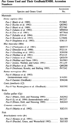
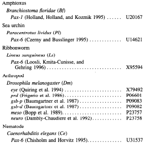
Results showed that the Pax gene family mainly has tissue-specific transcriptional controllers with a highly preserved DNA-binding area containing pairs with six alpha-helices. The research’s evolutionary analysis indicated that the genes in vertebrates (human and mouse) consist of four distinct and statistically supported clusters (Balczarek et al., 1997). The researchers argue that the collections arose before the deviation and evolution of vertebrates and Drosophila before the Cambrian radiation of triploblastic metazoan body strategies. The data obtained from the analysis is shown in the figure below (Balczarek et al., 1997).


The article “Evolution of Paired Domains: Isolation and Sequencing of Jellyfish and Hydra Pax Genes Related to Pax-5 and Pax-6” by Sun et al. (1997) examined a Pax-6 homolog and linked genes in cnidaria. Analysts argued that while other researchers have only investigated vertebrates as Walther et al. (1991), more primitive organisms could also contain a Pax-6 homolog that controls their eye development.
That is why Sun et al. (1997) chose two primitive cnidarians, a jellyfish and a hydra, as model organisms. Researchers conducted several key experiments, including isolation, and characterization of genomic clones, PCR amplification of paired box fragments, cDNA cloning of hydra paired box sequences, and electrophoretic gel-shift assay to find the evolutionary shifts in Pax genes in primitive organisms.
Results from the study indicated that hydra Pax-B contains both octapeptide and homeodomain, while Pax-A contains neither. Sun et al. (1997) argued that over time, there had been changes in Pax genes’ composition, which has led to the changes of Pax 5 and Pax 6. Phylogenetic analysis showed that Pax-B and Pax-A were closely related, making Pax genes be classified into two main groups. Researchers concluded that modern Pax-2/5/8 and Pax-4/6 genes evolved from ancestral genes that have similar characteristics.
Conclusion
The studies discussed in the paper show the evolutionary changes of PAX6 genes in vertebrates and primitive organisms. Investigations highlighted that the paired box transcription factor, PAX6, regulates the development of two different eye kinds: the vertebrate single-lens eye and the Drosophila compound eye. The results in the context of developmental biology mean that single genes, and the whole gene networks, retain similar functions across species and cause mutations related to human diseases, such as aniridia in the case of PAX6.
The results emphasize the fundamental principles of developmental processes, such as creating morphological diversity by remodeling existing information that the PAX6 gene does in the eyes, nose, and CNS. Some future directions in the area might be related to studies that discuss other organ’s development (nose, brain) and regulation in the embryonic development under the PAX6 activity and mutations of the gene in different vertebrates that can be prevented, if possible.
References
Balczarek, K. A., Lai, Z. C., & Kumar, S. (1997). Evolution of functional diversification of the paired box (Pax) DNA-binding domains. Molecular Biology and Evolution, 14(8), 829−842. Web.
Grindley, J. C., Davidson, D. R., & Hill, R. E. (1995). The role of Pax-6 in eye and nasal development. Development, 121(5), 1433−1442.
Sun, H., Rodin, A., Zhou, Y., Dickinson, D. P., Harper, D. E., Hewett-Emmett, D., & Li, W. H. (1997). Evolution of paired domains: Isolation and sequencing of jellyfish and hydra Pax genes related to Pax-5 and Pax-6.Proceedings of the National Academy of Sciences, 94(10), 5156−5161. Web.
Walther, C., & Gruss, P. (1991). Pax-6, a murine paired box gene, is expressed in the developing CNS. Development, 113(4), 1435−1449.
Walther, C., GuENET, J. L., Simon, D., Deutsch, U., Jostes, B., Goulding, M. D., Plachov, D., Balling, R., & Gruss, P. (1991). Pax: A murine multigene family of paired box-containing genes. Genomics, 11(2), 424−434. Web.