Introduction
A skeletal muscle cell is an essential component of contraction and represents the base unit of contraction. Normally a muscle contraction begins with an action potential from the CNS through an alpha motor neuron, which is then responsible for transmitting an action potential down its own axon to the motor endplate resulting in the release of the neurotransmitter acetylcholine, which binds to the acetylcholine receptors on the motor endplate activating them and opening its intrinsic sodium/potassium channel, causing an influx of sodium and an efflux of potassium, triggering an action potential.
Once this action potential is triggered, it normally leads to the opening of voltage-gated calcium channels leading to an influx of calcium as well as its release from the sarcoplasmic reticulum. The calcium then binds to Troponin C, found on actin-containing filaments. The Troponin causes exposure of the active binding sites by allosterically modulating Tropomyosin. Myosin then binds to the exposed site on actin resulting in a contraction which is modulated by ATP binding to the Myosin, releasing an inorganic phosphate and ADP. Therefore, lack of ATP results in a rigorous state characteristic of Rigor Mortis. While all the steps above occur, calcium is actively being pumped back into the sarcoplasmic reticulum. Thus, when there is no more calcium on the filament, Tropomyosin blocks the binding sites once again, causing a cease in contractions.
The mechanisms of sliding of the filaments are demonstrated in the diagram below.

A frog can easily be used as a skeletal muscle model as it contains muscle cells that are similar to most of the other vertebrates. This is especially in light of the fact that they are striated as the skeletal muscle of the human being, and since it is a vertebrate that does engage in a lot of muscle activity, especially its gastrocnemius muscle, which is greatly involved in its movement, it has a high amount of muscle fiber.
Materials and Methods
Experimental Preparation
- Cut the trunk away from above one of the femur bones, and then cut the muscles away from the femur.
- Place one blade of your scissors between the Achilles’ tendon (just above the heel) and the gastrocnemius muscle. Loosen the tendon by rotating the scissor blade around it. Cut across the flat distal portion of the tendon and tie a moistened thread across the tendon. After obtaining the muscle, the specimen immerse it into Ringer’s solution for about a minute before clamping the femur in a position above the writing pen and attach a thread to the pen.
- Fine copper wire can be used as electrodes. Obtain two pieces of copper wire (60 cm each) and scrape the ends with fine emery paper. One end of each copper wire should be inserted into the muscle, while the other end is inserted in the positive or negative end of the stimulator.
Experimental Protocol
Twitch Contraction
In order to stimulate the muscle and obtain a twitch contraction, we started with the following settings on the stimulator:
Frequency = 1/s
Delay = lowest
Duration = 0.1 ms
Voltage = lowest
The stimulator was then set for repeat mode, and the voltage gradually increased to obtain a contraction which we then recorded. The stimulator was then switched to single-mode, after which we measured and analyzed the tracings for the latent period, contraction period, relaxation period, and the recorded height of the contraction.
Summations
Quantal
The following settings were applied and the results recorded:
Frequency = 0.2/s
Delay = lowest
Duration = 0.1 ms
Voltage = lowest
The kymograph was set at the lowest speed, and the voltage gradually increased until contractions began. This was done until there were no further increments in the heights of contraction. (This was then graphed)
Temporal
The same setup above was employed with the kymograph speed at 1cm/s. The voltage level that caused maximal contraction was used, and the frequency was set at 1/second, and stimulation was done for 4s using the repeat mode; after allowing the preparation to rest for about 20s, the frequency was increased to 2/s, and the procedure was repeated for frequencies 4, 8, 16, 32, 64, and 128. The results were then compared.
The Effect of Load on Muscle, both Stretched and Unstretched
The muscle preparation was attached to the S hook, and the after-load screw was adjusted in order to bring about tension in the thread connecting the muscle to the lever, thus result in the bulk of the load being shouldered by the screw. The stimulus was then applied, and recordings were made.
For a stretched muscle, the same settings were applied except that the lever was adjusted to a free-loaded position.
Results
Twitch Contraction
The height of contraction at a threshold of 5.44 volts was 0.6 cm, with the contraction period lasting 0.0503 seconds and the relaxation period being 0.1353 seconds, and a latency period of 0.0223 seconds.
Summations
Quantal
For Quantal (spatial) summation, the contractions started out far apart and separated, and as the frequency increased, the contractions got closer and closer together until the line became flat and came back down. The data for temporal summation was as follows:
Effects of Load
The effects of applying a load on an unstretched muscle resulted in great stretch distances, but the rate of stretch decreasing with an increase in the amount of load.
For an already stretched muscle, the initial stretch distance was much smaller than for an unstretched muscle.
Discussion
In the experiment testing for twitch contraction, it can be argued that these periods of latency, relaxation, and contraction display the different levels of calcium concentration, with it being at its lowest during the period of relaxation.
The experiments above achieved good results, with the number of excited muscle cells increasing with an increase in the amount of voltage applied (in the experiment testing for quantal summation). This thus demonstrating that different cells have different thresholds.
The temporal summation experiment quite successfully demonstrated the theory behind stimulating titanic kinds of contractions before the muscle relaxes and the effects of varying stimuli on the timing of the contractions.
The effects of the load experiment serve to demonstrate the limiting factors when it comes to stretching of a muscle as well as the effect of applying a load to an already stretched muscle.
Graphs
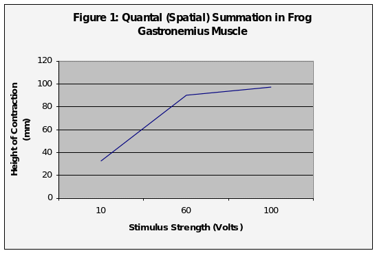
Figure one above shows the Quantal summation that is achieved by applying different levels of stimulus.
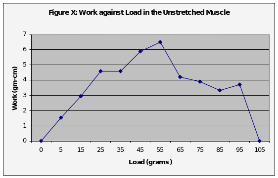
The figure above, figure 2 shows the amount of work against load in an unstretched muscle.
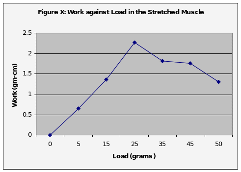
This figure depicts the amount of work against load in a stretched muscle.
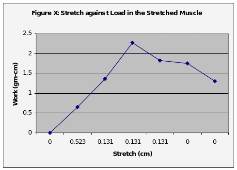
Graph number four shows the rate of a stretch against load in the stretched muscle.
References
Brooks, G.A; Fahey, T.D. & White, T.P. (1996), Exercise Physiology: Human Bioenergetics and Its Applications. (2nd ed.), Mayfield Publishing Co.