Introduction
Human beings live in the world through their senses. Without sensory receptors and the absolute threshold of seeing the external world we would not be able to see, we would know nothing about the world about us. In accordance to the above stated, seeing is as a result of perceived light, and the eyes are specifically configured to sense and receive light and colors. The absolute threshold of seeing is the smallest relative magnitude of stimulus energy needed to identify any stimulating information and its role is frequently described as the “access” or “passport” to the world.
For example, the minimum amount of light energy that makes it possible for a person to immediately notice an appearance of reflected light would be the absolute threshold for seeing that light. Though, the philosophical theory of seeing is believed to be produced by a special form of vitamin known as retinal. Retinal combines with another special protein called the opsin to form rhodopsin which is a known visual pigment.
The main work of rhodopsin is to transform the energy from light and the moment this energy is transformed from light, the nerve impulse is sent to the brain as explained by Gulley Ned (2011). This impulse allows an individual to see an image after it is formed in the eye through physiological processes which would be discussed in the subsequent paragraphs. This essay will explain the absolute threshold of seeing, and its physiological basis
Threshold for seeing and the physiology behind it
Absolute threshold of seeing are produced by the sense organs plus other parts of the body. The eye, for example, does not see all by itself but is a part of the entire visual network made up of nerves, muscles and the brain which, working together, produce sight. Although these complex networks use parts of the body to provide different kinds of sensations.
General characteristics
The general characteristic of vision is the absolute threshold of seeing. An individual cannot see all lights but will see a light when it reaches certain brightness. Absolute threshold is the smallest amount of physical stimulation needed to perceive any stimulating information or light. The absolute threshold is different for each individual; it will also change from time to time for the same person. For example, the absolute threshold for seeing a light may be higher or lower if an individual is exhausted or if the individual is relaxed. It will also vary if there are many other strong stimuli reaching the individual simultaneously. This means that the threshold for seeing is the smallest amount of energy needed to trigger vision in the rods and cones of the eyes.
Goldstein Bruce (2010, p.13) noted that there are three basic methods for determining absolute threshold namely:
- The methods of limits,
- The constant stimuli, and
- Adjustments
Michael Levine (2000, p. 100) also claims that the smallest amount of light energy that would enable a person to see a flash of light is the absolute threshold for seeing. However, Gelfand, 2004, gives the exact amount of light energy which is necessary to deduce vision. Gelfand claim that 90 photons of light energy are needed to deduce visual aspect of shone light which produces the ability to see. Furthermore, Gelfand asserted that only half of the light energy which is 45 photons enters the retina because the process in which information radiated energy is held occurs at the optic media.
Physiological basis for seeing
Before in-depth discussion on the physiological basis of seeing, it is imperative to discuss some of the important components which bring about the successful functioning of the eye. This is because we would dwell on the said anatomical components of the eyes as we discuss on the physiological factions of each which eventually brings about vision.
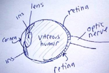
The human eyes are an amazing light sensing element. Their physical structure is astonishing. The eye consists of cornea, iris, retina, pupil, fovea, cones, rods, and optic nerve (see Figure 1) (Wandell 1995, p.63). In spite of the fact that they are interrelated, the eye’s functions are very explicit.
The cornea is an external, transparent enveloping structure that covers the eyeball. The iris is positioned below the cornea; it consists of an opaque diaphragm penetrated by the pupil. It is the iris that gives the eyes their pigment, a colorful mosaic imprint that-like the biometric identifications- is distinctive to each organism.
The black contractile within the iris is known as the pupil, and it determines the amount of light that goes through the eye’s lens- a transparent, almost sphere-shaped body that is causes visual focusing.
The retina is a wall of sensory receptors-rods and cones- on the backside of the eye. Rods, as shown by their name, are long, thin photoreceptors. Their extreme sensitivity makes them perfect for seeing unexpected movement-direct or peripheral- and for seeing in the dark. Human beings have more than 110 million rods within the retina. By comparison, the cones (which are cone-shaped and used almost solely for processing color) number around 7 million. Cones are used primarily for seeing during the day. Rods are used more for seeing at night or in dimly lit situations. The fovea, though small, is the most sensitive area of the retina. It consists only of cones, and it is exceptional at distinguishing colors and their subtle variations, sensing patterns of light and dark, and for noting meticulous detail.

The faculty through which the external world is visually apprehended probably affects human understanding. This is because the greatest proportion of information coming into the brains does so through the eyes.
The physiological basis of seeing has always been attractive. It is linked with creativity, learning, and perception. The physiological basis of seeing also plays a large part in biology, since it is relies on a triad of factors: the nature of light, the interaction of light and matter, and the physiology of human vision. Each element performs a fundamental role and their absence would make perception impossible.
Light is a form of energy known as electromagnetic radiation. Electromagnetic radiation consists of a large number of waves with varying frequencies and wavelengths. At one extreme are the radio waves having the longest wavelengths (several kilometers) and at the other extreme end are the gamma rays with the shortest wavelengths (0.1 nanometers). Out of the total electromagnetic spectrum a small range of waves cause sensations of light in the eyes. This is called the visible spectrum of waves.
The second part of the physiological basis of human vision is the retina. The retina is the photosensitive fraction of the eye and its superficial aspect consist photoreceptors. These perceives light and transmit it down through the optic nerve as an input to the brain. Additionally, the diverse frequencies give rise to the different color sensations in the eyes. Within the visible range, shorter and longer wavelengths give rise to different forms of seeing which can be passed through an optical device with a triangular shape used to invert a reflection.
The third factor is the interaction of light with matter. Whenever light waves strike an object, part of the light energy gets absorbed and/or transmitted, while the remaining part gets reflected back to the eyes.
Physiologically, electromagnetic radiation that produces visual sensation goes through the retina via the pupil and lens. The chemical interaction of the rods and cones stimulates the optic nerve, which transmits those image sensations to the brain. Whenever this happens, the electric signal passes from the photoreceptors (rods and cones) via a two neural chain which are the bipolar cells and then the ganglion cells before leaving retina through the optic nerve as the nerve impulses which are transmitted to the optic cortex. Durrant & Lovrinic (1984, p.30) confirm that the resultant occurrence of these processes is vision.
This function is achieved due to the distribution of the receptor cells over the entire retina- though not evenly – with only an exception of where the optic nerve (also known as the ganglion cell axons) leaves the eyeball. This spot is generally called the optic disc or the blind spot (Durrant & Lovrinic 1984).
The pathway of light
Primarily, whenever light passes from one object or substance to another, it differs from the previous substance in terms of density; subsequently the speed changes and its rays are bent or are termed as refracted (David 2005). The light rays bend in the cornea as they pass to aqueous humor, lens, then to vitreous humor. The electromagnetic radiation that produces visual sensation then contacts the innermost light-sensitive membrane covering the back wall of the eyeball where axons transmitting electrical discharge that travels along a nerve fibers from the retina are arranged at the rear part of the eyeball cells starting at the back of the eye as the cranial nerve that serves the retina. At the optic nerve, the several elongated, threadlike cells from the medial part of the eye interchange.
Gulley Ned (2011) asserted that this enables them to form the fiber tracts which are specially called the optic tracts. Each optic nerve is known to contain fibers from the lateral side of the eye on the same side and the medial side of the opposite eye (Gulley 2011). The optic fibers synapse with the neurons in the thalamus of the brain (which has axons from the optic radiation), and runs to the occipital lobe of the brain (part of the brain which integrates impulses for vision). Through the process of colligating with the cortical cells visual occurs (Goldstein 2010).
In brief, the light from the object which a person sees is physiological basis of vision. When the light enters the eyes, it goes through the cornea which represents the clear layer or covering of the eyes. Then it goes across the pupil which is a clear sphere in the iris. Immediately pupil senses the light, it enlarges (dilates) and controls the amount of light that passes through the eye’s lens. The lenses of the eyes then focus the light through the aqueous humor, which is a clear liquid that directs it to the retina (Wandell 1995, p.70).
The retina as a sensory receptors receives information and photos through its photoreceptors (rods and the cones), as previously stated. The optic nerves carry the information from the photoreceptors to the brain, and changed from the received form into a single image (Michael 2000). This process is performed through the release of neurotransmitters called the acetylcholine (in cones) and glutamate (in rods) (Goldstein 2010).
When rods are not activated, they are in the phase of depolarization. In this state they release a neurotransmitter called the glutamate. The process of loss of polarity is realized in the absence of light or illumination since the cells have a high intense mental effort of cyclic Guanine Monophosphate which is an energy rich compound, therefore inducing the path over which electrical signals can pass in the ion channels. Once the ion channels are opened, the sodium (K+) and Calcium ions (Ca2+) move through this space and stimulates the deficiency of electrons in the action as well as change of the cell which compels the discharge of the neurochemical that transmits nerve impulses across a synapse.
Immediately a gleam of light reaches the rods (as in cones), a special regulatory protein called the transducin is activated. Transducin brings about the physiological change of cyclic Guanine Monophosphate to Guanine Monophosphate through the activation of an enzyme called cGMP phosphodiesterase. This produces a feed-back mechanism which induces the prevention of the release of the neurotransmitters.
Importantly, when rods are open to a high intensity of electromagnetic radiation that produces a visual sensation for long, they turn out to be insensitive. This is the reason they are meant for night vision and built by the presence of Vitamin (Gulley 2011)
On the contrary, cones cells functions well in a high intensity of light, and generally called Photopic vision. Cones cells are mostly found on the region of the retina called the fovea (but are not evenly distributed in the retina). This is the section of retina where the retinal structures are separated, thus making it possible for electromagnetic radiation of visual sensation to fall without deviation on the visual receptor cell in the retina that is sensitive to bright light and to color. Gulley Ned (2011) explains that each cone synapses with a single interneuron which makes them to have greater visual acuity.
Conclusion
The absolute threshold of seeing, and its physiological basis is the least quantity of light energy that allows the perception of reflected light and the biological components that makes vision possible. Though, light is of great importance to seeing, and without it there would be no vision, because the retina senses light energy by the photoreceptor cells, the rods and the cones. David Rudd (2005) claimed that millions of receptor cells: the rods and the cones are found in the retina. The retina separates seeable messages into parallel neural processes that induce singular form of seeing. Besides, both rods and cones are called photoreceptors because they are the physiological basis of the eye which reacts to the electromagnetic radiation that produces visual sensation and make vision possible.
Reference List
David, RC 2005, Eye/Brain Physiology and Human Perception of External Reality. Web.
Durrant, D & Lovrinic, H 1984, Bases of Hearing Sciences. Second Edition. Williams & Wilkins, Owings Mills, MD.
Gelfand, SA 2004, Hearing an Introduction to Psychological and Physiological Acoustics. Fourth edition. Marcel Dekker. Inc., New York.
Goldstein, B 2010, Sensation and perception. Cengage Learning, Toebben Drive, KY
Gulley, N 2011, The Physiology of Seeing colors. Web.
Michael, L 2000, Fundamentals of Sensation and Perception, (3rd Edition). Oxford University Press, London.
Wandell, BA 1995, Foundations of Vision Science: Behavior, Neuroscience and Computation. Sinauer Associates Incorporated, Sunderland, MA.