Introduction/ Background
The liver is one of the largest organs in the human body. It is situated at the upper right-hand part of the abdominal cavity, underneath the diaphragm, and on top of the stomach, right kidney, and intestines (Westerhoff & Lamps, 2022). The liver has the shape of a cone, is dark reddish-brown in color, and weighs around three pounds. The organ is supplied with oxygenated blood by the hepatic artery and nutrient-rich blood by the hepatic portal vein (Westerhoff & Lamps, 2022). The liver holds approximately 13% of the body’s blood supply at any given time. The organ comprises two major lobes, which incorporate 8 segments that consist of 1,000 small lobes (Sagoo et al. 2018). The small lobes are joined to small ducts (tubes) that link up with larger ducts to form the common hepatic duct. The common hepatic duct transports the bile made by the liver cells to the gallbladder and duodenum through the common bile duct (Sagoo et al. 2018). This study intends to describe the gross and microscopic anatomy of the liver, its functions, and the diseases associated with it.
Gross Anatomy of the Liver
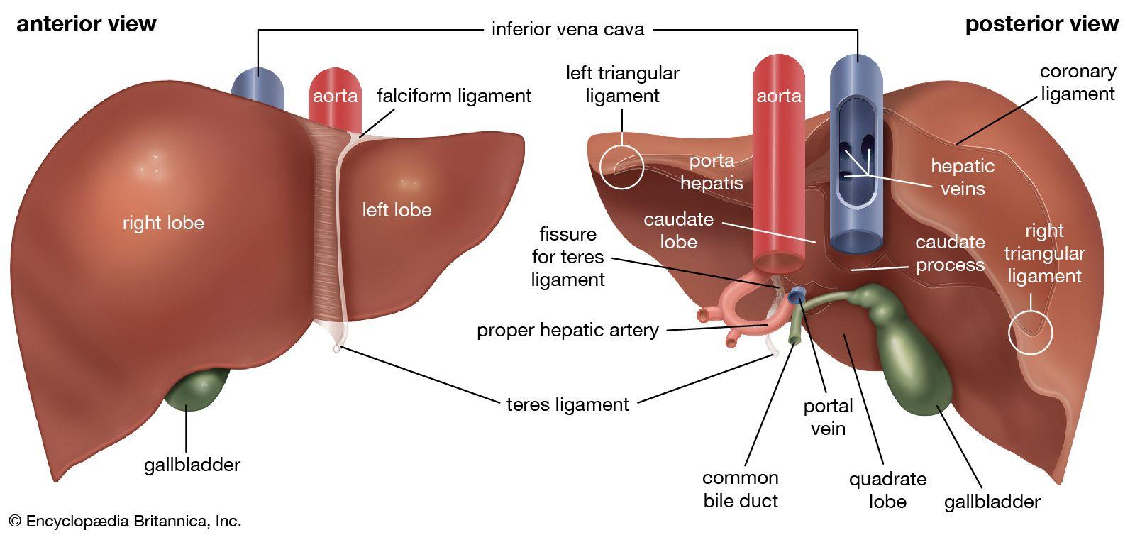
The liver is located beneath the lower right rib cage and takes more space in the upper right quadrant of the abdomen, with a segment spread out to the upper left quadrant. The organ is structured into two uneven lobes: a bigger right lobe and a smaller left lobe Frenkel et al. 2020). The left lobe is alienated on its anterior (frontal) surface by the thick falciform (sickle-shaped) muscle that joins the liver to the undersurface of the diaphragm (Frenkel et al. 2020). On the inferior surface of the organ, the right and left lobes are disconnected by a channel that contains the teres ligament, which goes to the navel. Two small lobes, the caudate and the quadrate, take a part of the inferior surface of the right lobe.
The liver regulates blood, which is supplied by two blood vessels. The hepatic artery, a major branch of the celiac axis, supplies the liver with fully oxygenated blood (Sagoo et al. 2018). On the other hand, the large portal vein, which acquires blood from all venous blood from the spleen, pancreas, gallbladder, lower esophagus, stomach, intestines, and part of the rectum, supplies the liver with partially oxygenated blood (Sagoo et al. 2018). At the porta hepatis, the portal vein splits into two bigger outlets, each entering one of the main lobes of the liver. Additionally, the porta hepatis is the departure point for the hepatic ducts (Frenkel et al. 2020). The above-discussed passages are the ultimate channels for a structure of smaller bile ductules interspersed across the liver that transport freshly manufactured bile from liver cells to the small intestine via the biliary tract.

Microscopic Anatomy of the Liver
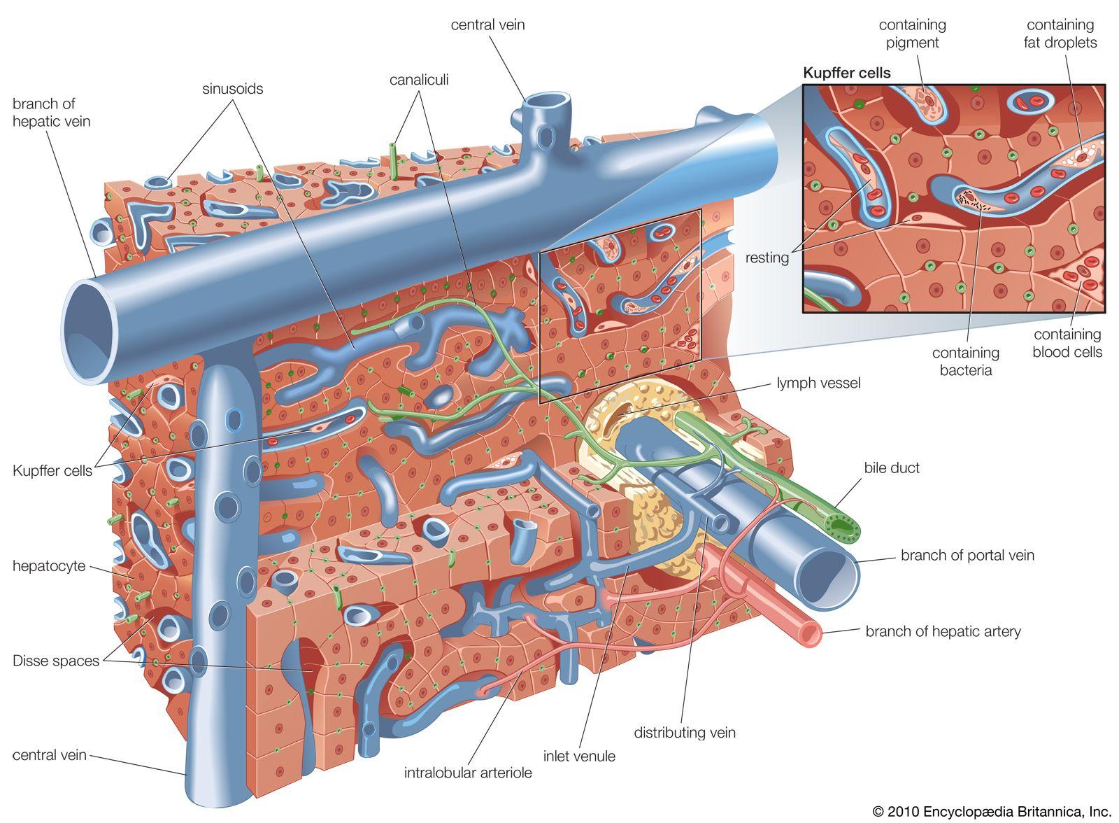
The microscopic anatomy of the liver demonstrates a unified configuration of collections of cells known as lobules, where the essential roles of the liver are conducted. Every lobule measures approximately one millimeter in diameter and comprises various cords of rectangular liver cells or hepatocytes (Westerhoff & Lamps, 2022). The cords radiate from central veins, or terminal hepatic venules, toward a thin layer of connective soft tissue that splits the lobule from other near lobules. The cords of liver cells are one-cell dense and are alienated from each other by numerous surfaces by spaces known as sinusoids or hepatic capillaries (Westerhoff & Lamps, 2022). Sinusoids are creased by thin endothelial cells containing outlets via which microvilli of the hepatocytes prolong, countenancing through ease of access of the hepatocyte to the bloodstream in the sinusoids.
The other major cell of the liver, the Kupffer cell, sticks to the surface of the sinusoid and ventures into its lumen. It acts as a phagocyte which is a cell that surrounds and destroys unwanted substances or cells. There are small spaces (Disse spaces) placed between the hepatocyte and the sinusoidal endothelium for the transportation of lymph (Sagoo et al. 2018). On neighboring exteriors, the hepatocytes are connected by thick, fitted intersections. The intersections are gone through by small pathways known as canaliculi, which are the terminal outposts of the biliary structure, getting bile from the hepatocyte (Sagoo et al. 2018). The canaliculi finally connect with other canaliculi to form increasingly bigger bile ducts that ultimately come out of the porta hepatis as the hepatic duct.

Functions (Physiology) of the liver
The main function of the liver is to regulate chemical levels in the blood and emits a substance known as bile. The liver produces bile which is utilized in carrying away waste products from the liver. All the blood from the stomach and intestines must go through the liver for regulation. The liver processes the incoming blood and breaks down balances, and creates nutrients. The organ further digests medicines or other drugs into forms that are easier to absorb in the rest of the body (Moradi et al., 2020). The liver is one of the body organs with many different functions. The most popular functions of the liver include detoxification, metabolism of carbohydrates, proteins, lipids, and hormones, and storing nutrients such as iron and vitamins (Moradi et al., 2020). The liver also acts as a synthesis of coagulation factors, production of bile, filtration, and storage of blood and therefore, its failure can lead to diseases.
Diseases of the Liver
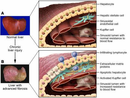
Liver fibrosis is one of the major diseases of the liver that result from a damaged liver. Liver fibrosis is the massive buildup of extracellular matrix proteins comprising collagen that happens in many kinds of chronic liver illnesses (Roehlen et al., 2020). Advanced liver fibrosis leads to cirrhosis, liver failure, and portal hypertension and often requires liver transplantation. In severe cases, if one does not have a liver transplant it results in death. The major causes of liver fibrosis include chronic hepatitis C virus infection, alcohol abuse, and nonalcoholic steatohepatitis (Northup, 2020). The accumulation of extracellular matrix proteins destroys the hepatic structure by creating a fibrous scar, and the consequent formation of nodules of regenerating hepatocytes describes cirrhosis (Roehlen et al., 2020). Cirrhosis leads to hepatocellular malfunction and augmented intrahepatic resistance to blood flow, which causes hepatic deficiency and portal hypertension.

Liver diseases are hard to detect in the early stages because their symptoms are similar to signs in other illnesses. Symptoms of liver disease include loss of appetite, inability to think, fluid buildup in the legs or stomach, jaundice, nausea, weight loss, and weakness (Northup, 2020). Since liver damage is caused by other issues, the treatment for liver fibrosis depends on the management of the underlying causes. For instance, if the cause is alcohol, a doctor can recommend a rehabilitative program to stop alcoholism. If the cause is non-alcoholic fatty liver disease, a doctor might suggest diet changes to lose weight.
References
Britannica, E. (2019). The Editors of Encyclopaedia Britannica. de la Enciclopedia Británica. Web.
El-Baz, L. M. F. (2014). Comparative Experimental Study between Mesenchymal and Hematopoietic Stem Cells in Rat Liver Regeneration (Doctoral dissertation, Suez University). Web.
Frenkel, N. C., Poghosyan, S., Verheem, A., Padera, T. P., Rinkes, I. H., Kranenburg, O., & Hagendoorn, J. (2020). Liver lymphatic drainage patterns follow segmental anatomy in a murine model. Scientific reports, 10(1), 1-9. Web.
Moradi, E., Jalili-Firoozinezhad, S., & Solati-Hashjin, M. (2020). Microfluidic organ-on-a-chip models of human liver tissue. Acta biomaterialia, 116, 67-83. Web.
Northup, P. (2020). History of coagulopathy in liver disease: legends and myths. Clinical Liver Disease, 16(Suppl 1), 56. Web.
Roehlen, N., Crouchet, E., & Baumert, T. F. (2020). Liver fibrosis: mechanistic concepts and therapeutic perspectives. Cells, 9(4), 875. Web.
Sagoo, M.G., Aland, R.C. & Gosden, E. (2018). Morphology and morphometry of the caudate lobe of the liver in two populations. Anat Sci Int 93, 48–57. Web.
Westerhoff, M., & Lamps, L. (2022). Liver: anatomy, microscopic structure, and cell types. Yamada’s Textbook of Gastroenterology, 157-167. Web.