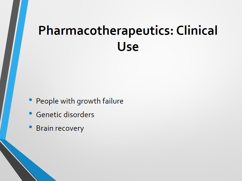
Anabolic Steroids and Testosterone Production Research Paper

Exclusively available on Available only on IvyPanda® Made by Human No AI

Introduction

Growth hormone (GH) fuels the development of skeletal and other tissues both directly and indirectly. Isaacs, Gardner and Baxter allege, “The development promotion actions of growth hormone are ‘indirect’ and are mainly mediated through generation of the insulin-like growth factor 1, which act both as an endocrine and paracrine hormone”(2023). The direct actions of the growth hormone are usually opposed to insulin. Nevertheless, there are severe insulin-like impacts of tentative physiological importance. Scientists allege that there is a significant correlation between growth, nutrition, and actions of growth hormones. The human pituitary secretes growth hormone in a pulsatile way. The pituitary gland has a distinct structural relationship with the hypothalamus. As a result, the anterior pituitary receives blood only from a portal venous system that empties the hypothalamus. In this regard, the hypothalamus regulates the secretion of the anterior pituitary hormone by injecting several factors into the circulation. The dynamic balance between the stimulatory and inhibitory hypothalamic somatostatin, peptides, and other hormones determines the release of growth hormones (Frantz and Rabkin 1474). This paper will discuss the neuroendocrine moderation of growth hormone release.

Anatomy and Physiology of Pituitary Gland

According to doctors, the weight of an adult pituitary is about 500g. The gland has a diameter of between 1.2 and 1.5 centimeters and it covers about 80% of the sellar space. The infundibulum connects the Pituitary gland to the hypothalamus. The gland is located at the base of the skull and enclosed by sphenoid bones. Having a picture of the physical structure of the pituitary gland can help one to learn the methods of bleeding and infarction in the gland (Frantz and Rabkin 1479). The pituitary gland constitutes of posterior and frontal (anterior) lobes linked to a central, midway portion as shown in the figure below.

Anatomy and Physiology of Pituitary Gland
Source: (Ross and Buchanan 145)

The anterior portion is also referred to as the adenohypophysis. The node is responsible for the production and release of six hormones, which are helpful in metabolic functions. The posterior lobe or what is scientifically known as neurohypophysis manufactures and releases two hormones. They are oxytocin and antidiuretic hormones. The posterior portion originates from the hypothalamus in the course of fetal growth. A piece of vascular tissue referred to as pars intermedia disconnects the anterior and posterior lobes. Initially, it was believed that the pars intermedia serves no purpose in the body. Nevertheless, some scientists claim that it helps in the production of hormone signals as well as the small quantity of the melanocyte-exciting hormone. The figure below represents the molecular structure of growth hormones.

Anatomy and Physiology of Pituitary Gland
Source: (Ross and Buchanan 147)

Pharmacodynamics and Pharmacokinetics of Growth Hormone

Growth Hormone and Plasma Protein

Scientists are yet to understand the physiological function of growth hormone-binding protein (GHBP). Nevertheless, it is believed that both the GHBP and growth hone receptor originate from the same gene. Terry and Martin hold, “Growth hormone and plasma protein form a complex at a one-to-two ration” (624). Yamashita and Melmed allege that there are two isoforms of plasma proteins in the blood (1011). “One of the isoforms binds the growth hormone with high affinity while the other binds with low affinity” (Yamashita and Melmed 1012). Scientists believe that the circulating plasma protein combines the growth hormone with superior affinity than the receptors. It does not matter the nature of the isoform involved. Research shows that plasma proteins with Exon 3 are firmly attracted to growth hormone. Additionally, the study indicates that the plasma carrier protein protracts the half-life of the growth hormone. The plasma protein does this by preventing growth hormones from fastening to growth hormone receptor.

Degradation and Metabolism

The nutritional state of people helps to regulate the level of growth hormone. Therefore, one can reduce the amount of growth hormone by consuming products that are rich in GH suppressors. Growth hormone helps to regulate the glucose level in the body. The increase in the degree of sugar in the body degrades growth hormone. Metabolic factors also degrade growth hormone. Intravenous or oral administration of glucose helps to degrade growth hormone secretion. Moreover, “insulin has a direct suppressive effect on growth hormone synthesis by pituitary cells in vitro through inhibition of growth hormone gene transcription” (Yamashita and Melmed 1011). The increase in the concentration of fatty acid in the body also degrades growth hormone. The hormone does not last for a long time in the body. After a few minutes, the growth hormone is transferred to the liver for metabolism. The liver converts growth hormone into numerous growth factors. One of the metabolic end-products of growth hormone is the insulin-like growth factor 1 (IGF-1).

Half-Life of Growth Hormone

Scientists allege that the production of human growth home ceases after puberty years. It underlines the reason many scientists produce synthetic growth hormone for use in adults. The major reason is the short-term half-life of growth hormone. Human growth hormone is not synthetic. As a result, it has a short half-life. Researchers hold that the half-life of human growth hormone ranges between three and four hours. The liver and kidney play a significant role in the metabolic clearance of the growth hormone. The two converts growth hormone into other products like insulin-like growth factor 1. It explains the reason people suffering from the hepatic disorder and renal failure show abnormalities in plasma growth hormone.

Regulation of Growth Hormone

Physiological Discharge

Growth hormone is discharged in form of successive pulses. The pulses occur after every three to four hours. Scientists claim that growth home is highly secreted when one is asleep (Quabbe, Schilling and Helge 1174). Mostly, the hormone is secreted at around 4 am before a person wakes up. The doctors claim that this is the period when counter-regulation is at the peak. The age of an individual influences the rate of the growth hormone pulsations. They are high at teenage, and decline as one grows old. Exercise, stress, sleep, and post-prandial reduction of sugar in the blood serve as stimuli to growth hormone discharge. Nevertheless, the scientists state that a majority of the incidents of secretion are impulsive (Terry and Martin 623). Doctors claim that there is a correlation between slow-wave sleep and the release of growth hormone. However, they are yet to substantiate this assertion as many samples of growth hormone discharged during sleep demonstrate significant heterogeneity. Indeed, the time when one is relax is what contributes to the increase in growth hormone secretion. Terry and Martin suggest, “Growth hormone discharge is a sleep-related event which probably should not be considered as closely linked to, or caused by, the neural process that subserves slow-wave sleep” (624). Growth hormone is one of the counter-regulatory hormones and is produced in high amounts mainly at night. Adrenalin, cortisol, and growth hormone alert the body when the level of insulin goes down. In other words, growth hormone offsets the impacts of insulin in the body. The increase in growth hormone leads to increase in blood sugar and vice versa.

Negative Feedback

There is significant corroboration that growth hormone regulates its discharge. However, it is not clear if the growth hormone regulates itself through direct or indirect actions. The growth hormone reaction to pharmacological incentives like insulin-stimulate arginine and hypoglycemia as well as physiological inducements like exercise and siesta is proved through pretreatment with human growth hormone (Mendelson, Jacobs and Gillin 487). Scientists argue that the attenuation to reaction to both physiological and pharmacological incentives could signify a direct action of the growth hormone. The growth hormone puts forth feedback at the pituitary and hypothalamic levels. Alteration of the growth hormone-releasing hormone (GHRH) and somatostatin (SMS) release regulates the secretion of growth hormone at the hypothalamus level.

Pretreatment of growth hormone inhibits the reaction to GHRH, particularly after an extended treatment that results in the rise in insulin-like growth factor 1 (IGF-1) levels. The growth hormone can then control its release autonomous of circulating insulin-like growth factor 1. The mechanism for this process may need the local production of insulin-like growth factor 1 in the pituitary or hypothalamus. The circulating insulin-like growth factor 1 also influences the release of growth hormone. Experiments conducted on human being show that growth hormone feedback takes place under the hypothalamic influence through somatostatin. Pyridostigmine, which is an acetylcholinesterase inhibitor that minimizes somatostatin secretion, obstructs growth hormone feedback. Additionally, there is adequate data from vitro research in the mouse, which show that insulin-like growth factor 1 and growth hormone induce secretion of hypothalamic somatostatin (Tannenbaum, Guyda and Posner 78). In the human being, “a recurring administration of growth hormone-releasing hormone at two-hourly intervals is accompanied by attenuation of the growth hormone response” (Salmon and Daughaday 834). However, constant administration of GHRH results in the increase in rate and amplitude of growth hormone release pulses. The increase occurs because the rate of growth hormone pulse depends on changes in background somatostatin activity. In return, the throb of growth hormone leads to the reduction of additional growth hormone secretion through the feedback loop.

Regulation by other Hormones

Other hormones regulate the secretion of growth hormone. They include steroid and thyroid hormones. Medical practitioners associate hypothyroidism in a human being with reduced growth hormone secretion. Children who suffer from severe hypothyroidism exhibit a drop in growth hormone pulse. Moderation of growth hormone is clearly delineated in vitro systems or animal models. The growth hormone gene of a mouse constitutes glucocorticoid and thyroid hormone-receptive features that contribute to increasing growth hormone generation (Gick and Bancroft 1989). Glucocorticoids can have constructive or destructive impacts on growth hormone production based on whether the principal effect is on GHRH and hypothalamic somatostatin generation or the pituitary somatotroph. In man, temporary usage of steroids results in increase in the secretion of growth hormone. On the other hand, long-term usage of the same suppresses the secretion of growth hormone (Frantz and Rabkin 1475).

Oestrogens contribute to the disparities in growth hormone secretion between men and women, in particular in relation to the increase in the release of growth hormone witnessed during teenage years. Oestrogens, both administered and endogenous boost induced and basal growth hormone secretion. Terry and Martin posit, “Women have higher basal levels of growth hormone than men, particularly during the high oestrogenic phase of the menstrual cycle” (624).

Physiological Conditions

The study indicates that physiological conditions like starvation influence the secretion of growth hormone. A study of a person who opted to fast for 40 days showed that his level of growth hormone increased from 0.73 to 9.86 (Ross and Buchanan 161). The level went up without the person taking drugs. Scholars claim that food suppresses the production of growth hormone. When one eats, the level of glucose goes high, therefore minimizing the secretion of growth hormone. Hunger stimulates the release of growth hormone. The body starts to burn stored fats, rather than to consume the lean mass (bone and muscle). The process of burning the fat stored in the body leads to increase in secretion of growth hormone. The time of the day also affects the release of growth hormone. Secretion of growth hormone is high in the early morning and normal during the day.

A study by Frohman and Jansson showed that healthy adults who went for five days without food recorded an increase in growth hormone pulse rate (232). Besides, they recorded high interpulse growth hormone levels as well as amplitude. The rise in pulse rate and amplitude was apparent on the first day that the subjects went without food. The reaction of the growth hormone to growth hormone-releasing hormone did not change throughout the fasting duration. The observation led to a conclusion that variations in pituitary sensitivity to GHRH do not result in changes in growth hormone release. A similar increase in growth hormone release upon food limitation has been recorded in numerous genus studied. Starvation and fasting contribute to low insulin-like growth factor 1 levels. Refeeding studies show that both protein and energy are vital in the generation of insulin-like growth factor 1. Anorexia also contributes to increasing the secretion of growth hormone.

Malnutrition and catabolic states enhance the discharge of growth hormone. The two lead to the augmentation of the amplitude of the growth hormone pulse and interpulse intensities. Acquiring the knowledge of pathophysiology of catabolic states and starvation can go a long way towards enhancing the management of such conditions. Doctors are yet to determine if insulin-like growth factor 1 or growth hormone can help to boost the nutritional status of sick individuals. The altered connection between IGF-1 and GH can serve as a defensive response in the emaciated patients. In the emaciated state, hypoglycemia is a constant threat, and necessary substrates are needed to assist the liver in manufacturing essential proteins. A reduction in insulin-like growth factor 1 may be a significant tolerant occurrence to facilitate muscle catabolism to produce metabolic fuels and avert the threat of hypoglycemia. In the same way, the additional increase in hormone growth intensities results in lipolysis and insulin resistance. Both can serve as defensive metabolic transformations in the event of severe and extended dietary control.

Receptors for Growth Hormone

The receptors for growth hormone were first illustrated in the rabbit liver. Tsushima and Friesen were the first to describe the cellular receptors for growth hormone. Today, scientists have identified the receptors for growth hormone in numerous vertebrate cell-types like adipocytes, chondrocytes, hepatocytes, and fibroblasts among others. Receptors for growth hormone occur as “disulphide-linked oligomers of a sialoglycoprotein-bidding subunit, which can be rapidly cleaved to lower molecular weight forms” (Isaacs, Gardner and Baxter 2024). As a result, receptors for growth hormone have a short half-life that lasts for about 45 minutes. Even though the receptors seem to be subject to transient down-regulation by growth hormone, the lasting impact of growth hormone is possibly receptor induction. Pregnancy and insulin activate the receptor while renal dearth and fasting minimize receptor expression.

Diabetes

Unlike the normal subjects, “the hyperglycemia of diabetes mellitus and sick patients with insulin resistance is associated with augmented rather than impaired growth hormone secretion” (Salmon and Daughaday 829). Individuals suffering from insulin-reliant diabetics exhibit higher rate and amplitude of growth hormone pulses. Moreover, they record high interpulse growth hormone intensities. The aberration of growth hormone discharge witnessed in diabetic patients seems to be inversely related to diabetic control. In diabetics and children suffering from malnutrition, oral administration of glucose does not trigger the ordinary containment of growth hormone discharge. Therefore, the nature of growth hormone discharge witnessed in diabetics is akin to that recorded in people who go without meals. The pattern of release signifies the impacts of intracellular hunger under the background of insulin paucity.

Obesity

Individuals suffering from obesity show reserved growth hormone reactions to many incentives including growth hormone-releasing hormone and insulin-provoked hypoglycemia (Ross and Buchanan 157). Notwithstanding this condition, children suffering from obesity have a tendency of growing at a fair rate. The levels of circulating insulin-like growth factor 1are elevated in people with obesity. In return, they affect the reaction of growth hormone to GHRH. However, this does not apply to all groups of people suffering from obesity. Once an individual loses weight, the growth hormone reaction to GHRH stabilizes. It shows that the change in GH response is as a result of the health condition and it does not cause obesity.

Growth hormone has both lipolytic and nitrogen-conserving actions. Therefore, growth hormone might facilitate weight loss among the patients suffering from obesity. Research shows that temporary inducement of growth hormone helps to minimize the wearing off of lean body mass among individuals who are fasting. On the other hand, it does not intensify fat loss. The same study has also shown that the dietary carbohydrate content influences sensitivity to growth hormone among patients suffering from obesity, and who do not feed on energy-rich products.

Conclusion

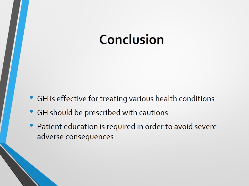
Growth hormone facilitates the development of skeletal and other tissues in the body. The functions of the growth hormone are connected to nutrition and growth. Pituitary gland manufactures the growth hormone. The hormone is bound by growth hormone-binding protein. Two isoforms of plasma protein facilitate the binding of growth hormone. The hormone does not stay for a long time in the body. The liver assists in the metabolic degradation of growth hormone. One of the metabolic end-products of growth hormone is insulin-like growth factor 1. The discharge of growth hormone is high at night. Sleep and post-prandial reduction of sugar in the blood augment growth hormone discharge. Health conditions like diabetics and obesity regulate the release of growth hormone. The discharge rate is high among people suffering from obesity and diabetics.

Works Cited

Frantz, Andrew and Mitchell Rabkin. “Effects of Estrogen and Sex Difference on Secretion of Human Growth Hormone.” Journal of Clinical Endocrinology and Metabolism 25.2 (2001): 1470-1480. Print.

Frohman, Lawrence and Jo Jansson. “Growth Hormone-Releasing Hormone.” Endocrine Reviews 7.3 (2002): 223-253. Print.

Gick, Gregory and Carter Bancroft. “Glucocorticoid Stimulation of Growth Hormone Massager Ribonucleic Acid Levels in GH Cells Inhibited by Calcium but not by Somatostatin.” Endocrinology 120.4 (2000): 1986-1990. Print.

Isaacs, Ramirez, David Gardner and James Baxter. “Insulin Regulation of Rat Growth Hormone Gene Expression.” Endocrinology 120.2 (2003): 2022-2028. Print.

Mendelson, Wallace, Laurence Jacobs and Christian Gillin. “Negative Feedback Suppression of Sleep-Related Growth Hormone Secretion.” Journal of Clinical Endocrinology and Metabolism 56.1 (2001): 486-488. Print.

Quabbe, Hummel, Erich Schilling and Heineken Helge. “Pattern of Growth Hormone Secretion during a 24-Hour Fast in Normal Adults.” Journal of Clinical Endocrinology and Metabolism 26.3 (1999): 1173-1177. Print.

Ross, Richard and Charles Buchanan. “Growth Hormone Secretion: It’s Regulation and the Influence of Nutritional Factors.” Nutritional Research Reviews 3.1 (1990): 143-162. Print.

Salmon, William and William Daughaday. “Hormonally Controlled Serum Factor which Stimulates Sulfate Incorporation by Cartilage in Vitro.” Journal of Laboratory and Clinical Medicine 49.6 (2001): 825-836. Print.

Tannenbaum, Gloria, Harvey Guyda and Barry Posner. “Insulin-Like Growth Factors: A Role in Growth Hormone Negative Feedback and Body Weight Regulation via Brain.” Science 220.4 (2004): 77-79. Print.

Terry, Leon and Robert Martin. “The Effects of Lateral Hypothalamic-Medial Forebrain Stimulation and Somatostatin Antiserum on Pulsatile Growth Hormone Secretion in Freely Behaving Rats: Evidence for a Dual Regulatory Mechanism.” Endocrinology 109.1 (1999): 622-627. Print.

Yamashita, Shunichi and Shlomo Melmed. “Insulin Regulation of Rat Growth Hormone Gene Transcription.” Journal of Clinical Investigation 78.1 (2000): 1008-1014. Print.

More related papers Related Essay Examples
Cite This paper
You're welcome to use this sample in your assignment. Be sure to cite it correctly

Reference

IvyPanda. (2022, June 10). Anabolic Steroids and Testosterone Production. https://ivypanda.com/essays/anabolic-steroids-and-testosterone-production/

Work Cited

"Anabolic Steroids and Testosterone Production." IvyPanda, 10 June 2022, ivypanda.com/essays/anabolic-steroids-and-testosterone-production/.

References

IvyPanda. (2022) 'Anabolic Steroids and Testosterone Production'. 10 June.

References

IvyPanda. 2022. "Anabolic Steroids and Testosterone Production." June 10, 2022. https://ivypanda.com/essays/anabolic-steroids-and-testosterone-production/.

1. IvyPanda. "Anabolic Steroids and Testosterone Production." June 10, 2022. https://ivypanda.com/essays/anabolic-steroids-and-testosterone-production/.


Bibliography


IvyPanda. "Anabolic Steroids and Testosterone Production." June 10, 2022. https://ivypanda.com/essays/anabolic-steroids-and-testosterone-production/.

If, for any reason, you believe that this content should not be published on our website, please request its removal.
Updated:
This academic paper example has been carefully picked, checked and refined by our editorial team.
No AI was involved: only quilified experts contributed.
You are free to use it for the following purposes:
  • To find inspiration for your paper and overcome writer’s block
  • As a source of information (ensure proper referencing)
  • As a template for you assignment
1 / 1