Introduction
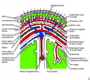
The scalp is the protective covering of skin and subcutaneous tissue surrounding the skull. The scalp covers the whole head, from the external occipital protuberance and upper nuchal lines to the lateral canthal and occipital edges (Al Bradford et al. 2019). Skin, connective tissue, epicranial aponeurosis, loose areolar tissue, and pericranium are the five layers that make up the scalp (see illustration below). The first three layers are permanently attached to the scalp. The scalp begins at the supraorbital ridges and continues through the external occipital protuberance and superior nuchal lines.

The scalp protects the cranial vault from injury and infectious microorganisms by acting as a physical barrier. The scalp’s aesthetic value extends beyond its functional role as a physical shield. Connecting the occipitofrontalis tendon is a thick layer of solid fibrous tissue known as the galea aponeurotica or epicranial aponeurosis (Bradford et al. 2019) Its tight attachment to the thick subcutaneous layer of connective tissue may stop the scalp from being stretched, which is very helpful during surgery. The flexibility of the scalp relies on this loose connective tissue. It’s not just a pretty face; it’s a movable plane that divides the trigeminal from the pericranial (Seery, & Gerard 2022). The pericranium is the most profound layer of the scalp and is made up of thick, uneven connective tissue (Almulhim et al. 2019). It has a strong bond with the calvarial bone in the skull. Due to the blood vessels found there, the underlying calvarium is well-supported.
Embryology
The ectoderm forms the embryo’s outermost layer of tissue. The notochord initiates an event that partitions the neural ectoderm from the surface ectoderm. Separate from the exterior ectoderm, which develops into the epidermis, a neural tube and crest emerge, giving rise to the nervous system (Polak‐Witka et al. 2020). Furthermore, during gastrulation, mesoderm is formed from the ectoderm. The scalp’s skin, or epidermis, develops from the mesoderm. In contrast, the scalp’s connective tissue, or dermis, originates from the mesoderm—a congenital disability known as aplasia cutis genetic causes a patch of skin to be missing at birth. The area is most often the crown of the head, although other areas of the body, such as the trunk and limbs, are not immune. The epidermis may be affected directly, or the dermis, bone, or dura may be missing in a localized area. However, aplasia cutis congenital is considered to be linked to chromosomal abnormalities, teratogens, intrauterine issues, thrombotic events, and trauma.
Nerves
From the ophthalmic branch of the trigeminal nerve, the frontal nerve branches out to become the supratrochlear and supraorbital nerves. These nerves carry the vast bulk of the sensations felt on the front of the head. The zygomaticotemporal nerve, which arises from the maxillary branch of the trigeminal nerve, and the auriculotemporal nerve, which arises from the mandibular branch of the trigeminal nerve, give lateral sensory innervation. The front of the head is innervated by the minor and greater occipital nerves. The medial tactile innervation of the scalp behind the ear is carried out by the medial spinal nerve, which has its source in the cervical plexus. The larger spinal nerve, which originates from the cervical spinal nerve 2 dorsal rami, innervates the posterior scalp in anterosuperior direction toward the scalp’s crown.
Conclusion
The frontalis belly is a muscle that starts in the galea aponeurotica and ends where your eyebrow does. Originating from the mastoid and superior nuchal lines, the occipitalis muscle travels posteriorly to intrude on the galea aponeurotica (Malmande et al. 2018). The superior zygomatic nerve and posterior auricular nerve provide the nerve endings that activate these muscles. Together, they may raise the brows and tuck the scalp. When the frontalis muscle contracts, it causes the skin on the forehead creases (Sokoya 2018). It’s essential to observe that the layer of slack connective tissue goes all the way to the muscles. Black eyes, or periorbital hematomas, result from bleeding into this area, which may go down to the orbicularis oculi.
References
Almulhim, Abdulaziz M., and Mohammed Madadin. 2019. Scalp laceration. StatPearls Publishing.
Bradford, Benjamin D., and Judy W. Lee. 2019. “Reconstruction of the Forehead and Scalp.” Facial Plastic Surgery Clinics 27(1): 85-94.
Malmande, Vithal, Naveen Rao, Amaresh Biradar, Abhilash Bansal, and Chandrika Dutt. 2018. “Scalp replantation in a cervical spine injury patient: Lessons learnt.” Indian Journal of Plastic Surgery 51(2): 243-246.
Polak‐Witka, Katarzyna, Lidia Rudnicka, Ulrike Blume‐Peytavi, and Annika Vogt. 2020. “The role of the microbiome in scalp hair follicle biology and disease.” Experimental Dermatology 29(3): 286-294.
Seery, Gerard E. 2022. “Surgical anatomy of the scalp.” Dermatologic surgery 28(7): 581-587.
Sokoya, Mofiyinfolu, Jared Inman, and Yadranko Ducic. 2018. “Scalp and forehead reconstruction.” In Seminars in Plastic Surgery 32(2): 090-094.