Introduction
A cell is the basic functional, structural, and biological unit of a living organism (Latham 2009). The element can reproduce independently. As such, cells are regarded as the building blocks of life. Cells are made up of cytoplasm, which is held within a membrane. In addition, they contain numerous bio-molecules.
In this paper, the author will analyze animal cells, tissues, and primary organ systems. The report will cover the structure and functions of animal cells and organelles. It will also address the structure and functions of tissues and main organs of the body.
The Structure and Function of Animal Cells and Organelles
The Main Structural Features of an Animal Cell
Animal cells are eukaryotic. Thibodeau and Patton (2003) note that their structure varies based on their type and function in the body. However, a typical animal cell has various distinct structural features.
Table 1: The main structural features of an animal cell.
The Functions of the Main Parts of an Animal Cell
An animal cell is made up of a membrane-bound nucleus. Tortora and Nielsen (2012) note that the cell contains a number of cellular organelles. The numerous parts carry out distinct functions. The main components include the cell membrane and the extracellular matrix. Others are the nucleus, golgi apparatus, mitochondria, and endoplasmic reticulum. It also contains ribosome, lysosome, peroxisome, and cytoskeleton.
Table 2: The functions of the major parts of an animal cell.
The Structure and Sub-Types of Body Tissues
A tissue is a group of cells that have the same functions and shapes. Light (2004) notes there are four primary forms of tissues in humans. They include connective, nervous, epithelial, and muscular tissues. Each of these has a number of sub-tissues.
Connective Tissue
Overview
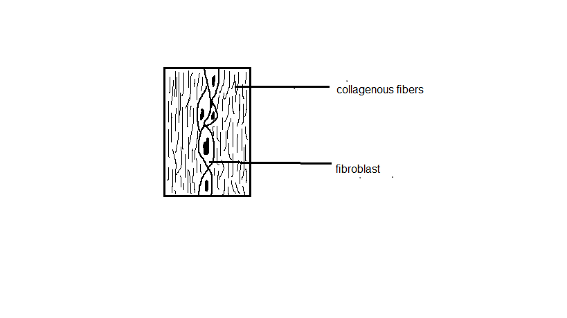
They are fibrous in nature. They comprise of cells separated by extracellular matrix. Connective tissues are made up of three primary components. The constituents are fibres, cells, and ground substances. However, Clancy (2011) observes that blood and lymph do not contain the three major elements.
The drawing below is a depiction of a connective tissue:
Subtypes of connective tissues
There are diverse sub-types of connective tissues with different structures. They include blood, bone, fibrous connective tissue, adipose tissue, and cartilage.
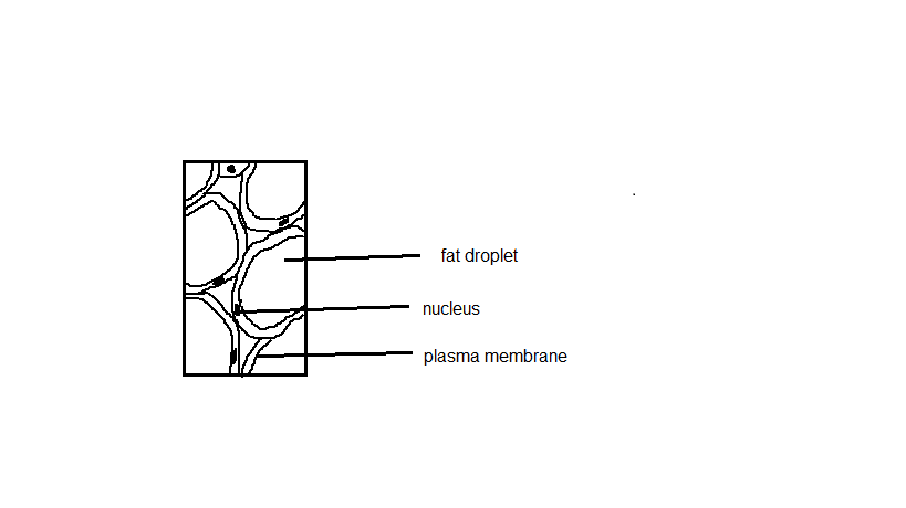
Adipose tissue is one of the most important sub-tissues. It is made up of 80% fat and adipocytes (Martini & Timmons 2006). It is regarded as a primary endocrine organ. The reason is that it secretes several hormones. Such hormones include estrogen, cytokine, leptin, and resistin. The figure below is an illustration of adipose tissue:
Cartilage
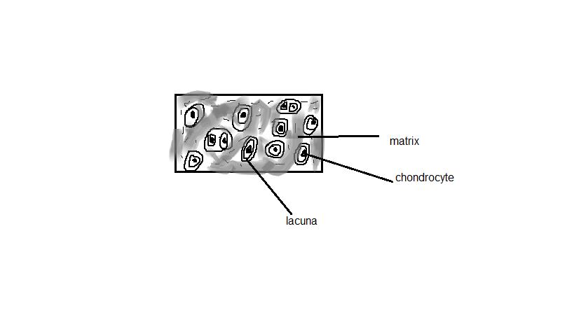
The subtype is made up of specialised cells referred to as chondroblasts. According to Marieb and Hoehn (2010), the cells produce copious amounts of ground substance and extracellular matrix. The figure below is a depiction of this sub-tissue:
Blood
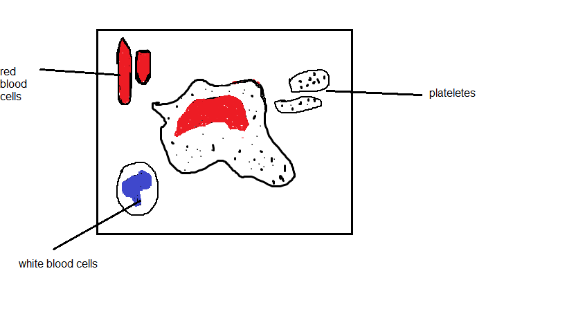
Blood is a special body fluid in animals. The subtype of connective tissue is made up of cells suspended in plasma. The figure below illustrates this connective tissue:
Epithelial Tissue
The tissue is formed by cells that cover the surfaces of different organs. Cells that make up the epithelial layer are joined by semi-permeable and rigid intersections (Manolis 2009).
Subtypes of epithelial tissue
There are numerous subtypes of epithelial tissues. They include simple squamous, cuboidal, columnar, stratified squamous, and stratified cuboidal tissues. Each subtype has a unique structure.
Simple squamous
They are thin, flat, and single layers tightly fit together. In addition, they are permeable and susceptible to damage. The figure below shows a simple squamous:
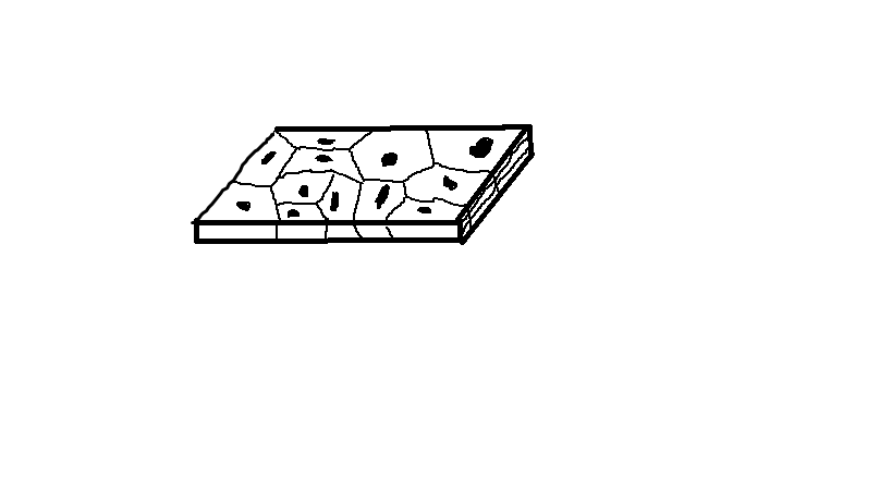
Simple Cuboidal
Fox (2011) notes that the epithelial subtypes are single layered and cube-shaped.
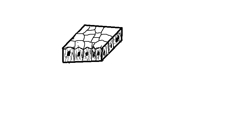
Simple columnar
The epithelial subtype is single layered, elongated, and nucleic (Mai 2012). In addition, it is comprised of microvillus and goblet cells.
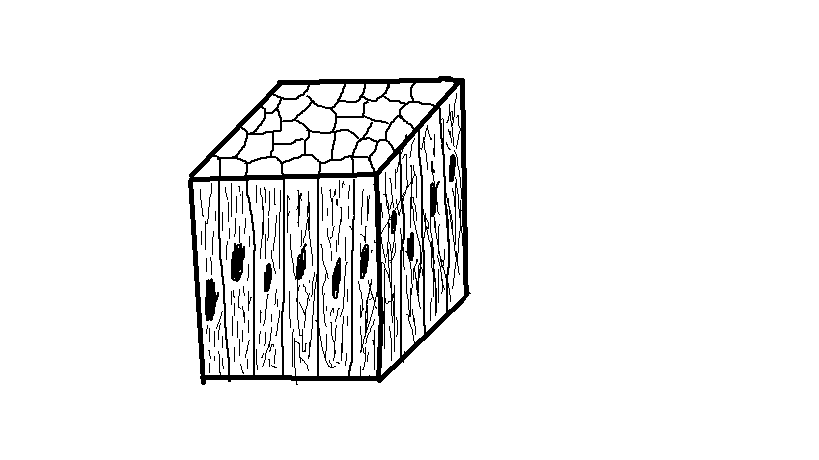
Nervous Tissue
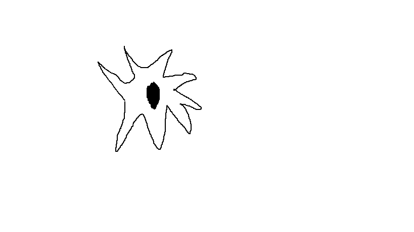
The nervous tissue is composed of specialised cells that receive stimuli and conduct pulses to and from all parts of the body. The cells making up the nervous tissues are long and string shaped.
Subtypes of nervous tissues
Neurons
They exist in different forms and sizes. According to Mai (2012), neurons are characterised anatomically. The structural classifications include unipolar, bipolar, multipolar, and anaxonic.
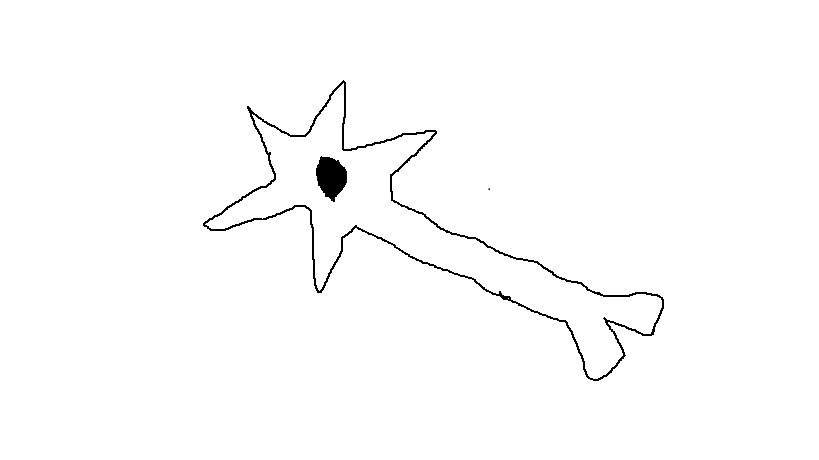
Glial cells
According to Latham (2009), these cells comprise of ample support systems. They ensure proper functioning of the nervous system.
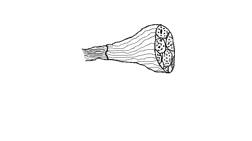
Muscle Tissue
A muscle tissue is made of excitable cells with contraction abilities. In addition, it contains lots of microfilaments, which consist of contractile proteins. The muscle tissue is the most common of all the four subtypes.
Subtypes of muscle tissues
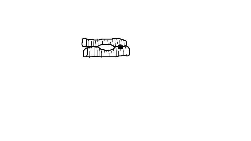
Cardiac muscle
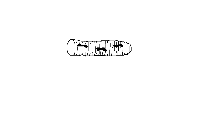
It is made up of cardiomyocyte cells, which contain three nuclei. In addition, it has numerous cross-striations. The striations are formed by revolving fragments of protein fibres (Mohrman & Heller 2010).
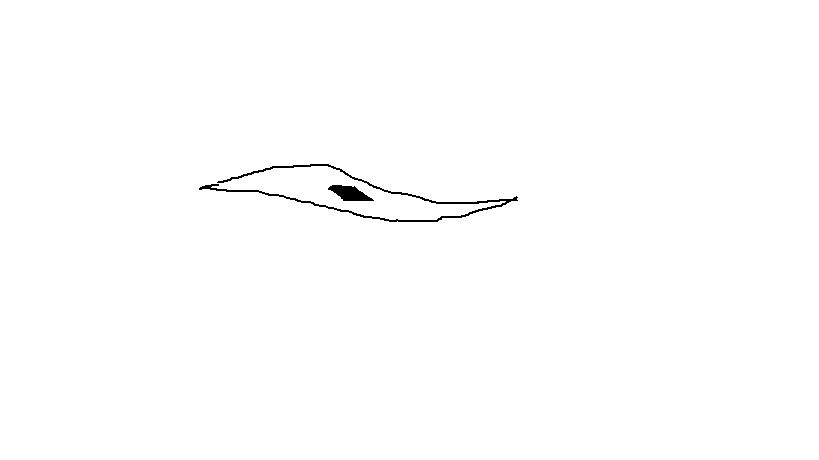
Smooth muscle
Smooth muscles have a fusiform shape and a tensing and relaxing capability. In a relaxed state, the tissue is often spindle-shaped.
Skeletal muscle
The muscles are made up of myocytes (Thibodeau & Patton 2003). They contain collagen fibres referred to as tendons. The fibres help them append to the bones.
The Location and Function of the Four Main Tissues and Sub-Tissues 1.1. Connective Tissue
The tissues are located between other tissues found in the animal body (Martini & Timmons 2006). They cover most animal parts. The functions of connective tissues include providing a medium of diffusion for nutrients and oxygen into the cells from the capillaries. In addition, they prevent the overstretching and tearing of organisms.
Connective tissue subtypes: Location and functions
Adipose tissue
The tissue is located in the liver and other muscles (Manolis 2009). Its primary purpose is to store energy in form of lipids. In addition, it stifles and insulates the body.
Cartilage tissue
The tissue is found in various parts of the animal. The parts include ribcage, nose, ear, bronchial tubes, knee, and between the bones. Other areas are elbow and intervertebral discs. The main function of cartilage is to connect organs.
Blood
It is found in the entire body. Its primary purpose is to transport nutrients and oxygen to the cells (Clancy 2011). In addition, the component is responsible for carrying away metabolic waste.
Epithelial Tissue
The tissue is found in the skin. Its primary function is to cover surfaces found inside the organism and body cavities.
Epithelial tissue: Location and function of subtypes
Simple squamous
They are located in the walls of capillaries, air sacs, and blood vessels. Their primary function is filtration and diffusion (Marieb & Wilhelm 2014).
Simple Cuboidal
They are located in the kidney tubules, liver, salivary glands, and pancreas. Their main function is secretion and absorption.
Simple columnar
They are found in the lining of the digestive tract and uterus (Thibodeau & Patton 2003). Their primary function is the secretion of such substances like mucus and enzymes.
Nerve Tissues
The tissues are located in the brain, heart, lungs, and kidneys. Their primary functions include controlling, supporting, and protecting neurons.
Nerve tissues: Location and function of subtypes
Neurons
They are found in the brain and the spinal cord. The primary function of neurons is the transmission of information (Carter & Aldridge 2009).
Glial cells
They are located in the brain and the spinal cord. Their main function is to insulate neurons.
Muscle Tissues
The tissues are located in the heart, bones, and hollow organs. Their main function is to facilitate the movement of the organ walls and the skeleton.
Location and function of muscle sub-tissues
Cardiac muscles
They are located in the heart. Their primary function is to synchronize the heartbeat.
Skeletal muscles
They are found in the bones. They are intended to move bones (Manolis 2009).
Smooth muscles
They are located in the arteries, digestive tract, and bladder. Their primary function is to facilitate movement in hollow organs.
Location, Size, and Function of the Main Organs of the Body
Table 3: Main organs of the body.
Three Major Organs with Reference to Tissue Types and Subtypes
Table 4: Three major organs.
The table above shows three main organs with reference to their tissues, sub-tissues, and system.
References
Carter, R & Aldridge, S 2009, The human brain book, DK Pub., London.
Clancy, J 2011, The human body close-up, Firefly Books: Richmond Hill, Ontario.
Fox, S 2011, Human physiology, 12th edn, McGraw-Hill, New York.
Fried, G & Hademenos, G 2013, Biology, 4th edn, McGraw-Hill Education, New York.
Latham, D 2009, Cells, tissues, and organs, Raintree, Chicago, Illinois.
Light, D 2004, Cells, tissues, and skin, Chelsea House, Philadelphia.
Mai, J 2012, The human nervous system, 3rd edn, Elsevier Academic Press, Amsterdam.
Manolis, K 2009, The skeletal system, Bellwether Media, Minneapolis.
Marieb, E & Hoehn, K 2010, Human anatomy & physiology, 8th edn, Benjamin Cummings, San Francisco.
Marieb, E & Wilhelm, P 2014, Human anatomy, 7th edn, Pearson, Boston.
Martini, F & Timmons, M 2006, Human anatomy, 5th edn, Pearson/Benjamin Cummings, San Francisco.
Mohrman, D & Heller, L 2010, Cardiovascular physiology, 7th edn, McGraw-Hill Medical, New York.
Thibodeau, G & Patton, K 2003, Anatomy & physiology, 5th edn, Mosby, St. Louis.
Tortora, G & Nielsen, M 2012, Principles of human anatomy, 12th edn, John Wiley & Sons, Hoboken.