Medial Malleolus Injury
For this question, I chose chip or avulsion injury of the medial malleolus. Chip fracture entails one type of medial malleolus injury caused by the rapture of a ligament. Chip fractures occur when the ligament cuts from the inside of the ankle, which then pulls the bone that the ligament attaches to, causing bone breakage. In most chip fractures, the bone is typically attached to the ruptured ligament, and can sometimes, such injuries can be associated with simple ankle sprains (Claveau & Fallat, 2020). While the injury might appear simple, the presence of a chip or an avulsion fracture might point out the presence of a more serious injury.
Injury Dynamics of Chip Fractures
The injury dynamics for an avulsion injury depend on the type of ligament that rapture around the medial malleolus. A patient might suffer from anterior medial malleolus avulsion injury due to the rapturing of the anterior tibiotalar ligaments that connect the distal tibia to the talus bone of the foot. Anterior medial malleolus avulsion can also occur when the tibionavicular ligament breaks lose. The second type of medial malleolus injury entails the lateral surface of the distal tibia, which is connected to the calcaneus by the tibiocalcaneal ligament. When this ligament cuts in cases of trauma in sports or manual work, the surface of the medial malleolus may come off, resulting in bone injury (Carter et al., 2019). Lastly, posterior medial malleolus avulsion injury can result when posterior tibiotalar ligaments break, making the back of the medial malleolus to be lost.
Functional and Structural Losses that Exist with Chip Injuries
The medial malleolus is an important anatomical point of the leg as various important structures pass posterior to it. Behind the medial malleolus are the tibial nerve, flexor hallucis longus tendon, flexor digitorum longus tendon, and posterior tibial artery. Since medial malleolus injury entails cutting part of the bone in the distal tibia, the cut bone might have sharp edges that could cut the adjacent structures. For instance, the posterior tibia artery might be cut by the sharp bone edges leading to a structural loss. As a consequence of this artery damage, the plantar part of the leg will not receive blood, as the posterior tibial artery that divides into medial and lateral plantar arteries will be open (Zhao et al., 2020). The presentation of cut arteries is profuse bleeding where the patient might show a hematoma if the bleeding is covered by a membrane or an open wound if the sharp edges of the bone damage even the skin.
Along with the arterial injury, nerve injury is the next common manifestation of both structural and functional loss. The sharp edges of the broken medial malleolus may cut the tibial nerve as the nerve is soft. This damage prevents nerve impulses from reaching medial and lateral plantar nerves, which are the main divisions of the tibial nerve (Claveau & Fallat, 2020). As a result, muscles such as flexor hallucis brevis, abductor hallucis, and flexor digitorum brevis will not be supplied, leading to difficulty and pain during gait and other toe deformities.
Compensation with Gait Cycle for Chip Injuries
Walking with a broken medial malleolus can be difficult and can affect the position of one’s gait. To walk on an avulsion fracture, one needs to ensure that the level of comfortability is maximum before putting weight on the injured ankle. Nevertheless, the gait of such patients is typically compensated by the use of crutches, especially in the early stages (Zhao et al., 2020). Additionally, the gait can be restored if the movement of the broken ankle is minimized using a cast, which increases the chances of recovery.
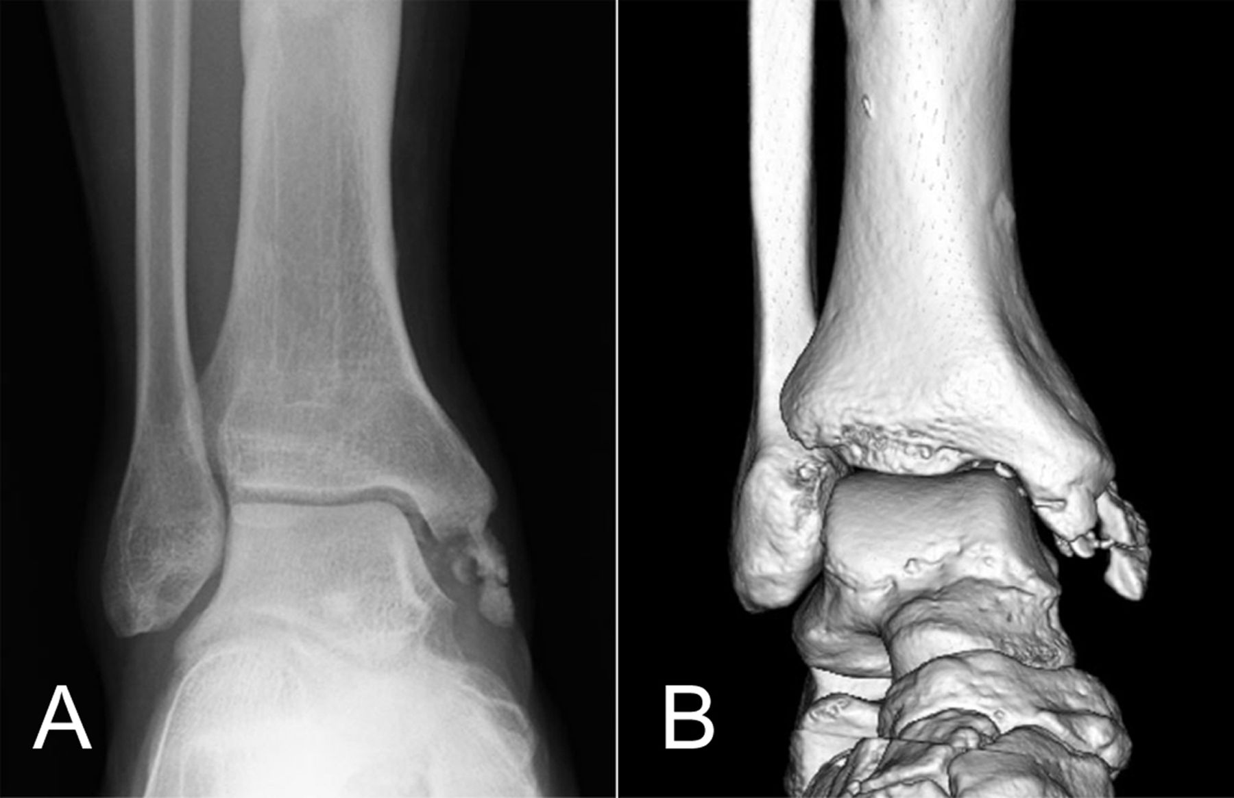
Medial Malleolus Chip Injury Image

The photo above is divided into one for an X-ray and the other for a musculoskeletal ultrasound. Both images represent a medial malleolus avulsion injury where the lower part of the medial malleolus has broken from the upper part. From the picture, one can see the medial malleolus being in pieces resulting from the avulsion fracture. By comparing the medial malleolus to the lateral malleolus, it is visible that the lateral malleolus is intact as opposed to the appearance of the former.
References
Carter, T. H., Duckworth, A. D., & White, T. O. (2019). Medial malleolar fractures: Current treatment concepts. The Bone & Joint Journal, 101(5), 512-521.
Claveau, T., & Fallat, L. M. (2020). An unusual case report of a stage IV osteochondral defect imitating a medial malleolar avulsion fracture. The Journal of Foot and Ankle Surgery, 59(3), 590-593.
Zhao, J., He, M., & Fang, Z. (2020). An exceptional avulsion fracture above the medial malleolus: A retrospective case series. Journal of the American Podiatric Medical Association, 110(5).