Background
Cholelithiasis is one of the most common gastrointestinal disorders experienced globally. It comes about when the lumen of the gall bladder is infested with stones or calculi (Chiang et al. 2008). Thus, it is sometimes referred to as gallstone disease. Cholelithiasis in general is a serious burden for healthcare systems. Statistics do indicate that an estimate of 700, 000 cholecystectomies are carried out in the United States, more than 190,000 in Germany, and more than 40,000 in Chile (Lammert and Miquel 2008, p.124). However, it has been found that the occurrence of gallstones increases with age and the female gender, with 20% of the patients being females and 5% being males all lying within the 50 to 65-year age group (Chae et al. 2004, p.162).
A briefcase history
At one point in time, a 47-years-old obese woman exhibited on and off epigastric pain for 4 months. The pain was naturally dull but sharpened after consumption of fatty meals. On Physical examination everything was normal; however, Laboratory investigations depicted that the liver functioned normally as well as the white cell count. It was, therefore, suggested that a clinical diagnosis of gallstone is made. An abdominal x-ray (AXR) was performed as a routine initial investigation (Ahuja et al. 2006, 360).
The other case involved, a 46-year-old obese woman who presented a 6-hour history of moderate pain in the RUQ that began after eating dinner. Her pain became more severe for a certain duration of time before becoming constant. The patient indicated that this was not the first time she had experienced such pains but had neglected to seek medical attention. Even though all her vital signs were normal, physical examination indicated some tenderness to palpation especially on the upper side of the right arm.
Clinical diagnosis
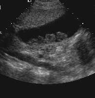
On clinical diagnosis, the patient complains of very serious and constant pain that began in the epigastric and regions of the umbilical cord before localizing in the right hypochondriac area. She further claims that the pain rotates around the chest and on the lower side of the inferior angle of the scapula. This is noticed due to the tenderness as well as some stiffness in the right hypochondriac region. She has high levels of white cells and a moderate fever. After the ultrasound, the results show the presence of several multiple stones in the gall bladder (Witmer, L. M. 2003).
Anatomy
The gallbladder abbreviated as GB is a sac taking the shape of a pear. It consists of three main parts, namely the fundus, neck and body. On the distal end of the gallbladder is the fundus which is very broad. The body forms the main part of the gallbladder with the neck being at the proximal end. The neck normally extends to the cystic duct which has a length of about three to four centimetres. In addition, the cystic duct comprises numerous membranous folds referred to as the spiral valve found alongside the duct. The folds play an important role in preventing the cystic duct from collapsing or distending.
In most cases, the gallbladder is about seven to ten centimetres in length with a width of three centimetres. The gallbladder can hold up to 30 to 40 millilitres of bile. Bile is a digestive juice produced by the liver in the small lobules and thereafter travels to either the right or left hepatic ducts which form the common hepatic duct via the small ducts. After its production, bile may either be temporarily stored in the cystic duct through the aid of the common bile duct; it is secreted into the duodenum.
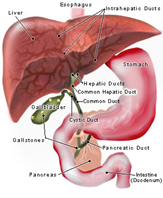
The common bile duct is normally positioned on the rear end of the superior part of the duodenum. The head of the pancreas fits into the descending part of the duodenum. Thus, the rears of the pancreatic and common bile ducts are closely related and given the name ducts of wiring. 40 per cent of human beings have the two ducts separated with each having separate openings into the duodenum. The remaining 60 per cent have the two ducts commonly joined thus having a common passage referred to as hepatopancreatic ampulla into the duodenum via the single papilla. In such individuals, the single-channel narrows down as it gets into the duodenum hence becoming prone to gallstones. This is as a result of the circular muscle fibre called the hepatopancreatic sphincter or the sphincter of Oddi which relaxes after an increase in levels of cholecystokinin (CCK) in the blood. When this happens, the resulting muscular ring induces a protrusion in the lumen of the duodenum. This prostration is referred to as the duodenal papilla (Bontrager and Lampignano 2005, p.527).
Pathology overview
Cholelithiasis or rather Gallstones is a pathological condition characterized by the presence of stones or calculi within the lumen of the gallbladder (Chiang et al. 2008). The gallstones are solid and are formed from the bile in the gallbladder. The biliary system functions to produce digestive enzymes and bile which aids in fat digestion. Produced by the liver, bile is made up of different substances such as cholesterol and bilirubin which is a waste product of the normal breakdown of blood cells in the liver. After production, bile is normally stored in the gallbladder to be used when the need arises. For instance, after consumption of a high-fat and high-cholesterol meal, the gall bladder is induced to contract thus with the help of the common bile duct it injects bile into the ileum. Once in the small intestine, bile assists in the digestive process. In most cases, gallstones are classified as cholesterol and pigment stones. More than 90 per cent of the gallstones found in the gallbladder are mainly made up of cholesterol (Lammert and Miquel 2008, 124).
There are two types of gallstones namely:
- Cholesterol stones- These arise from an excess of cholesterol in the bile. It is more common making up about 80 per cent of all the gallstones.
- Pigment stones – These are formed when bilirubin is in high quantities in the bile.
The size of gallstones varies from the tiny ones as grains to the very large ones as a golf ball. The smaller stones are a common occurrence some people may have only a single large stone or a combination of both (Lammert and Miquel 2008, p.124). The small stones normally form what is called sludge.
Symptoms of Cholelithiasis vary with the size and/or the number of the gallstones present. This is since the presence of gallstones in the gallbladder may cause no problems. However, large and many gallstones may lead to pain in the gallbladder especially in response to a fatty meal. In case the gallstones move out of the bladder, there might be instances of pain as a result of blockages of the ducts connecting to the pancreas, gallbladder, liver or small intestine. If this happens, there will be serious complications. The complications will be due to the entrapment of enzymes in the duct which causes infections, organ damage, and very severe pain and if left untreated may result in death. Gallstones are a common phenomenon in overweight middle-aged women although elderly men and women are also prone to the disorder. Research has also indicated that women who have at one time been pregnant have higher chances of developing gallstones compared to those who have never been pregnant (Balentine, J. R. 2009).
Role of imaging modalities in the diagnosis
Plain radiography
Often, gallstones occur in singular or multiple in the RUQ. This is evidenced using the US. Plain radiographs are only able to detect 50 per cent of the pigment stones and 20 per cent of the cholesterol stones (Goldschmid et al. 2006, p.291).
OCG
Using the OCG, it is possible to detect single or multiple luminously filled defects within a gallbladder that has been altered. This is because of their high dependency on gravity. The gallstones containing air or high cholesterol levels usually float and are mobile as depicted by the change in position of the films and supine. To send away the bowel gas, compression films are essential (Goldschmid et al. 2006, p.291).
Computed Tomography
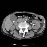
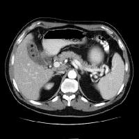
Detecting the presence of gallstones on CT is a difficult task. It is the same as using sonograms. CT is preferably used to further characterize the complications of the disorder rather than being used in the initial study. CT is of great help when it comes to the detection of intrahepatic stones or recurrent pyogenic cholangitis (Goldschmid et al. 2006, p.291).
Magnetic Resonance Imaging
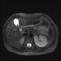
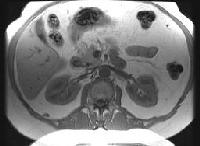
MRI is not used as a screening tool. Finding stones using the abdominal MRI is in most cases incidental. This is because the accuracy of identifying gallstones using the MRI has not been proven. To do this, confirmation findings with the US are required to rule out polyps or tumours. As a matter of fact, distinguishing gallstones from polyps or tumours is not an easy task. At times the gallstones may be overlooked due to their small size or as a result of the respiratory or motion artefacts (Goldschmid et al. 2006, 291).
Ultrasonography
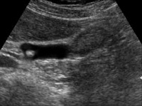
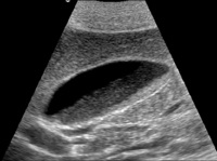
When using the US to examine the gall bladder, it is possible to see the whole organ from the distal end (fundus) to the common duct when the patient lies facing upwards. With ultrasound, the sensitivity and specificity are very high at 95 per cent since it is possible to identify gallstones as small as 2mm (Malik, Malet and Leonard 2004, p.326). In addition to this, sonography is preferred because of its simplicity, speed, safety in pregnancy and because it does not expose the patient to the effects of radiography (Chiang and Santen. 2008).
Treatment and prognosis
On diagnosis of the disorder, the patient has to undergo an operation to surgically remove the gallbladder. However, in instances where the gallstones have no symptoms, treatment is not essential (Balentine, J. R. 2009). In cases of acute Cholelithiasis, Laparoscopic cholecystectomy is the best option (Heidelbaugh, 2006, p.359).
For the surgery, small tube-like instruments are used to carry out the procedure. The instruments function to make a small opening on the abdomen through which the gallbladder is taken out. The instruments used are usually installed with a camera. The entire process is less painful compared to open surgery. In addition, it has minimal chances of causing complications as well as having a faster recovery period. The procedure is carried out by a general surgeon and takes a duration of 20 minutes to one hour while the patient is under anaesthesia.
In other cases, the procedure begins with laparoscopy and then the abdominal procedure follows. This happens when laparoscopic surgery is not workable for a specific person. The open procedure is commonly used in cases of biliary tract infection or scars from previous surgeries.
Once in a while, ERCP (Endoscopic Retrograde Cholangiopancreatography) is executed just before or during the surgery to find any gallstones that may have relocated from the gallbladder to other parts of the biliary system. Once detected, they are immediately removed to eliminate cases of recurrence in future.
ERCP also may be performed after surgery to determine the presence of gallstones in the biliary tract. In other cases, ERCP is carried out without surgery especially in people who are sick or too weak to undergo surgery (Balentine, J. R. 2009). Incidental findings of gallstones may need a follow up until symptoms are detected. Thus, surgical procedure asymptomatic gallstones with no complications are barred due to the risk of complications that could crop up as a result of the intervention. It is therefore important to have a close follow-up on diabetic patients and pregnant women who have asymptomatic gallstones as they are at great risk of complications (Strasberg, S. M. 1997, p.656-658).
Summary and conclusion
Cholelithiasis is one of the most common gastrointestinal disorders experienced globally which arises when the lumen of the gall bladder is infested with stones or calculi. This disorder could lead to death if left untreated. However, the recent technological developments in imaging modalities which include; computerized tomography, ultrasound, radiography and magnetic resonance have made the process of diagnosis simpler as well as treatment. Of all the mentioned modalities, Ultrasound is the choice of preference due to its high sensitivity, simplicity, speed and safety. All in all, this assignment has been of great help to me as I have gained a thorough understanding of the pathological disorder Cholelithiasis. The ability to easily detect the signs and symptoms as well as their demographics is a great step towards a faster diagnosis of Cholelithiasis disorder.
Reference List
Ahuja, A. T., G. E. Antonio, K. T. Wong, and H. Y. Yuen. (2006). Case Studies in Medical Imaging. Cambridge: Cambridge University Press.
Balentine, J. R. (2009). Gallstones.Web.
Bontrager, K. L., and J. P. Lampignano. (2005). Radiographic Positioning and Related Anatomy. St. Louis: Mosby, Inc.
Brunetti, J. C. (2009). Cholelithiasis. Web.
Chae, F. H., C. M. Robert, Jr, Md, V. S. Gregory, and E. Ben. (2004). Cholelithiasis (Gallstones). In Surgical Decision Making (Fifth Edition), 162-165. Philadelphia: W.B. Saunders. Web.
Chiang, W. K. (2008). Cholelithiasis: Treatment & Medication. Web.
Chiang, W.K., F. M. Lee, and S. Santen. (2008). Cholelithiasis. Web.
Goldschmid, S., b. Edited, D. B. Thomas, Md, L. W. Teresa, P. M. Michael, E. Consulting, and David, Z. (2006). Evaluation of the Specificity and Sensitivity of Biliary Imaging. In Zakim and Boyer’s Hepatology (Fifth Edition), 291- 296. Edinburgh: W.B. Saunders. Web.
Heidelbaugh, J. J. (2006). Right Upper Quadrant Abdominal Pain (Cholelithiasis). Essential Family Medicine (Third Edition), 359-365.
Philadelphia: W.B. Saunders. Lammert, F., and J.-F. Miquel. (2008). Gallstone disease: From genes to evidence-based therapy. Journal of Hepatology 48 (Supplement 1): S124-S135. Web.
Malik, A. H., P. F. Malet, and J. Leonard. (2004). Cholelithiasis, Complications of. In Encyclopaedia of Gastroenterology, 326-333. New York: Elsevier. Web.
Strasberg, S. M. (1997). Cholelithiasis and acute cholecystitis. Bailliére’s Clinical Gastroenterology 11 (4): 643-661. Web.
Witmer, L. M. (2003). Clinical Anatomy of the Biliary Apparatus: Relations &Variations. Web.