Trichuris trichiura is a nematode called ‘whipworm’ commonly present all over the world. It is thought to be carried by nearly one-quarter of the world’s population, with a prominent presence in tropical Asia followed by Africa and South America (Donkor, 2006). It is an intestinal helminth parasite with a comparatively smaller size than Ascaris lumbricoides (Donkor, 2006). It causes a disease called Trichuriasis (whipworm disease/Trichocephaliasis) (Material Safety, 2001).
Taxonomy
The whipworm can be classified as follows:
- Class: Adenophorea
- Subclass: Enoplia
- Order: Stichosomida (Trichurata)
- Superfamily: Trichocephaloidea
- Family: Trichuridae
The normal length of the adult worm is 30-45mm for males and 35-50mm for females. It preys on tissue secretions in the intestinal mucosa by burying its thin threadlike anterior half into it (Donkor, 2006).
Epidemiology
The whipworm is almost ubiquitous in its presence, with nearly one-quarter of the world’s population carrying it. It is most common in areas using untreated fecal matter as fertilizer (Material Safety, 2001). Hyperendemic areas may have a 90% infection rate (Material safety, 2001). Coming to the United States, it is rare as a whole, but with mild presence in the Southeast, infecting nearly 2.2 million people (Donkor, 2006).
Trichuriasis is not fatal generally. However, heavily infected people may suffer from rectal prolapsed. There is no racial predilection for the disease. Furthermore, children are mostly infected. The two main reasons for it are that children may consume soil either directly or indirectly and lack the initial immunity that comes with age (Donkor, 2006).
Pathophysiology
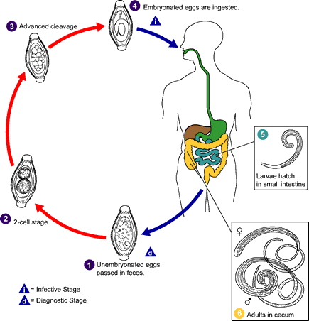
The eggs become infective after 10-14 days in soil. It is transmitted through the fecal-oral route. The incubation period depends on the infection load and nutritional status (Material safety, 2001). Unlike Ascaris lumbricoides and hookworm, whipworm does not have any tissue migration phase (Donkor, 2006). The larvae hatch in the intestine, and the adult worm resides in the large intestine, especially in the cecum and colon (Donkor, 20).
It usually takes three months from the ingestion of eggs to the development of adult worms. The female lays 2000-10000 eggs per day for a period of 5 years (Material safety, 2001; Donkor, 2006). The eggs are barrel-shaped with terminal polar plugs with an approximate size of 52 µm x 22 µm (Material safety, 2001). Coming to immunity, cytokines such as interleukin 25 (IL-25) that mediate type 2 immunity are necessary to control inflammation in the gastrointestinal tract (Donkor, 2006).
There is no direct person-to-person transmission of the infection. Generally, eggs can be observed in the feces 70-90 days after ingestion. Untreated carriers may shed eggs for years (Material safety, 2001).
Diagrammatic Representation of the Life Cycle
Clinical Manifestations
Patients are mostly asymptomatic. If the worm burden is less than ten worms, no symptoms usually appear (Parasitology, 2007). Children who eat dirt fall under the risk category.
The symptoms and signs can be described as follows:
Symptoms
- Nocturnal loose stools (Donkor, 2006).
- Chronic profuse mucus and bloody diarrhea may occur in patients with longtime infection or containing more than 200 worms (Parasitology, 2007; Donkor, 2006).
- Edematous rectal prolapsed in severe cases (Parasitology, 2007).
- Malnutrition, anemia, and weight loss
- Vague abdominal discomfort
- Growth retardation in children with chronic infection (Donkor, 2006).
Signs
- Mild to severe abdominal tenderness
- Signs of anemia
- Prolapsed rectum
- Characteristic clubbing of fingers
- Adult worms can be directly visualized on rectal mucosa by anoscopy or when the rectum is prolapsed (Donkor, 2006).
Diagnosis
It is made on the basis of symptoms and by detecting eggs in the feces (Parasitology, 2007).
Laboratory Investigations
The following features can be observed through laboratory studies.
- Eosinophilia can be observed due to progressive tissue invasion (Donkor, 2006).
- Findings indicative of anemia
- Oval-shaped eggs with transparent bipolar terminal plugs are seen on stool smear (Donkor, 2006). Hence, stool smear is advised to see eggs as well as parasites.
- Adult worms emerging from the bowel mucosa can be observed on proctoscopy (Donkor, 2006).
Differential Diagnosis
It includes chronic anemia, gastroenteritis, and giardiasis (Donkor, 2006). Proper diagnosis can be made through stool examination.
Management of Trichuriasis
No emergency management is necessary except in cases of rectal prolapsed and severe anemia.
Treatment
A 3-day treatment with mebendazole at a dose of 200mg for adults and 100mg for children is effective (Parasitology, 2007). A single dose of 500mg mebendazole results in a 40-75% cure rate (Donkor, 2006). Albendazole can be used as an alternative drug, though it is not as effective as mebendazole (Donkor, 2006).
The parasite biochemical pathways are adequately different from the host to allow chemotherapeutic management (Donkor, 2006). Mebendazole (Vermox) irreversibly blocks the uptake of glucose and other nutrients in the host intestine and selectively kills the worm (Donkor, 2006). It should be avoided in patients with hypersensitivity and pregnancy. Moreover, interaction with carbamazepine and phenytoin may decrease its efficiency, whereas cimetidine may increase it (Donkor, 2006). Precaution and dose adjustment are needed while using the drug in hepatic impairment cases.
All in all, proper prevention and management can completely eliminate the infection.
References
Donkor, K. A. P. (2006). Trichuris Trichiura. eMedicine from WebMD. Web.
Material Safety Data Sheet-Infectious Substances. (2001). Public Health Agency of Canada. Web.
Parasitology-Chapter four. (2007). In Microbiology and Immunology On-line: University of South Carolina School of Medicine. Web.