Abstract
It is often difficult for pathogens to penetrate the brain-blood barrier. However, some of them do. Toxoplasma Gondii, Naegleria Fowleri, and Taenia Solium are common pathogenic elements that could successfully penetrate this barrier. However, there is scanty medical literature explaining the mechanisms used by these pathogens to penetrate this blood-brain barrier. This paper bridges this research gap by giving more insight into how pathogens penetrate the barrier. However, the scope of this analysis is limited to the three pathogens mentioned – Toxoplasma Gondii, Naegleria Fowleri, and Taenia Solium. The research aims to find out if Naegleria Fowleri, Taenia Solium, and Toxoplasma Gondii use the same mechanism to cross the blood-brain barrier. A comprehensive assessment of existing research studies shows that Naegleria Fowleri, Taenia Solium, and Taxoplasma Gondii do not use the same mechanism to cross the blood-brain barrier. This assertion supports the main research hypothesis, which claims that Naegleria Fowleri, Taenia Solium, Taxoplasma Gondii use different mechanisms to cross the blood-brain barrier. Furthermore, this paper shows that pathogens use different mechanisms to minimize the effect of the host’s immune system.
Introduction
The blood-brain barrier plays an important function in the proper functioning of the central nervous system because it separates the blood system from the brain’s extracellular fluid (Velázquez-Moctezuma, Domínguez-Salazar, & Gómez-González, 2014). Indeed, for the central nervous system to work properly, the molecules passing across the blood-brain barrier are often tightly regulated (Manno, 2012). The blood-brain barrier does so by binding the cerebral endothelial cells together to prevent the flow of infectious molecules between the blood system and the brain (Velázquez-Moctezuma et al., 2014). The same mechanism prevents the diffusion of molecules between the endothelial cells and the brain (Pardridge, 2006). Masocha and Kristensson (2012) further explain this structural barrier by saying, “The cerebral endothelial cells have low levels of pinocytotic activity or transcytosis and form a functional barrier by selectively transporting only specific molecules into the brain parenchyma” (p. 202). This way, a key function of this permeable membrane is preventing parasites and pathogens from infiltrating brain tissues. Therefore, the blood-brain barrier only allows the selected permeability of molecules that are important to the human neurological function to pass the blood-brain barrier. It could also allow for the entry of water and specific gases into the brain tissue (Masocha & Kristensson, 2012). However, using different mechanisms, intracellular and extracellular parasites could still invade the central nervous system (Khan, 2008). Such infections could cause neurological damage or disturbances that could be fatal to victims. However, host-derived immune molecules fight such attempts. Nonetheless, some of these blood-borne pathogens have derived unique mechanisms for invading the blood-brain barrier, thereby undermining the host’s immune response (Davson, 1993). In light of this observation, Masocha and Kristensson (2012) says,
“The Th1 immune response, which is directed against intracellular pathogens, can be inhibited during infections with certain microbes in which the Th2 response, which is directed against extracellular pathogens, instead, is promoted; the two arms of the immune response being mutually inhibitory” (202).
Pathogenic infections to the brain are often rare. However, when they occur, they could cause serious damage to the brain. Indeed, as Masocha and Kristensson (2012) observe, parasitic infections in the brain usually have serious medical consequences. In fact, in most cases, they are fatal (Masocha & Kristensson, 2012). Such fatalities often occur because the parasites prevent antibodies from circulating in the brain, thereby causing disturbances in the brain cycle (Masocha & Kristensson, 2012). This is why many patients who suffer from parasitic infections also suffer from other conditions, such as seizures or epilepsy (Masocha & Kristensson, 2012). However, a patient’s immune system could help to minimize these effects.
Some common parasites have a propensity to invade the immune system. For example, Toxoplasma gondii, Naegleria Fowleri, and Taenia Solium have a strong propensity to invade the blood-brain barrier and sabotage the host’s immunity (Velázquez-Moctezuma et al., 2014; Roy et al., 2014). Some parasite-derived molecules may support the invasion of these blood-borne pathogens in the brain. Recent medical studies have explored how bacterial infections could occur through the blood-brain barrier (Khan, 2008). Most of their assessments have not specifically focused on how blood-borne pathogens could similarly infiltrate the central nervous system, but they have explored how various microbes spread across the system (Masocha & Kristensson, 2012). Therefore, there is little information known regarding how blood-borne pathogens cross the blood-brain barrier and cause such infections. There is also scanty information to explain how the immune system controls these parasites. To fill this research gap, this review explores the mechanisms used by Naegleria Fowleri, Taenia Solium, and Toxoplasma Gondii to cross the blood-brain barrier.
Comparison/Contrast
Mechanism of Naegleria Fowleri
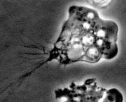
Naegleria Fowleri is among a few known parasites that affect the central nervous system and penetrate the blood-brain barrier (see figure 2) (Cardona, Restrepo, Jaramillo, & Teale, 1999). Naegleria Fowleri occurs naturally and causes primary amebic meningoecephalitis (PAM) (Chávez-Munguía et al., 2014). This condition has a high fatality rate of 97% (Masocha & Kristensson, 2012). Certain conditions cause this amoeba to thrive. For example, warm and moist environments aid its spread. Usually, these non-pathogenic elements are in the soil or water bodies. There is no clear explanation showing why Naegleria Fowleri targets the brain because its non-pathogenic nature makes it a less likely infiltrative element (Toney & Marciano-Cabra, 1994). However, its invasive mechanism stems from how amoeba enters its host. The common mode of exposure, for human beings, is swimming in freshwater bodies, which exposes them to the amoeba. Entry usually occurs through the nasal cavity. This point of entry allows it to attach to the nasal mucosa (Masocha & Kristensson, 2012). Later, it moves through the olfactory nerves and invades the brain tissue through the olfactory epithelium and the cribriform plate (Burri et al., 2012). After penetrating the brain tissue, Naegleria Fowleri destroys the central nervous system by penetrating through the brain vasculature (Masocha & Kristensson, 2012). This entry gives it access to the frontal lobes of the brain after reaching the meninges region. This infection mechanism greatly relies on adhesion because the amoeba has a surface protein that improves its bonding to fibronectin (Masocha & Kristensson, 2012; Burri et al., 2012). Researchers have always said that this surface protein is similar to the human integrin-like receptor (Chávez-Munguía et al., 2014). Nonetheless, the exceptional bonding with fibronectin becomes part of the matrix of the host’s cells. Naegleria Fowleri incapacitates the host’s immune system because it has efficient locomotive skills. Its relatively direct access to the site of infection also incapacitates the immune system from preventing an attack on the central nervous system (Chávez-Munguía et al., 2014).
How does it cope with the Immune System?
Few kinds of literature explain how CNS infective amoeba copes with the human immune system. However, there is enough medical evidence to show that Naegleria Fowleri has developed efficient mechanisms for evading the immune system. To explain how it does so, Stanford University (2015) says, “The amoeba attacks cells by tragocytosis and the release of a plethora of cytolitic enzymes, including aminopeptidases, hydrolases, esterases, acid, and alkaline phosphatases and dehydrogenases” (p. 7). The ability of Naegleria Fowleri to penetrate the blood-brain barrier also stems from its highly cytopathic effect. It contributes to the amoeba’s virulence by damaging impeding cells (Stanford University, 2015). Although the host’s immune system may produce cytolytic molecules to impede the destruction of the brain tissue, Naegleria Fowleri could defeat its attempts by creating a strong resistance to lysis (Chávez-Munguía et al., 2014). Regarding this analysis, Stanford University (2015) says, “Research suggests eukarytoic cells, such as mammalian erythrocytes, neutrophils, and tumor cells utilize complement-regulatory proteins to protect themselves from lysis by the complement system of the innate immune system” (p. 8). Naegleria Fowleri has also created strong resistance to the host’s immune system by adapting to carrying complementary regulatory proteins. The ability of Naegleria Fowleri to shed the membrane attack complexes also prevents the host’s immunity from destabilizing its infection mechanism (Chávez-Munguía et al., 2014). Based on these adaptive mechanisms, scientific research has yet to prove that the human immune system has any effect on Naegleria Fowleri’s attack mechanism (Stanford University, 2015). However, research has affirmed the presence of Naegleria Fowleri antibodies among people who suffer from amoeba exposure (Stanford University, 2015). However, there are usually a small number of these antibodies, thereby making it difficult to detect them during the pathogenic analysis (Burri et al., 2012). This failure leaves the host’s immune system as having the best mechanism to prevent infections by Naegleria Fowleri. However, the amoeba has developed elusive adaptations to evade its mechanisms, thereby leaving the host exposed to brain infections. However, this assessment does not mean that the host’s immune system is helpless to brain infections by Naegleria Fowleri because the amoeba has several vulnerabilities as well.
The Vulnerabilities of Naegleria Fowleri’s Invasive Mechanism
Although the above section of this paper shows the adaptations of Naegleria Fowleri, the amoeba is still vulnerable to some facets of the host’s innate immune system. This vulnerability comes from the amoeba’s weakness to neutrophils. The host’s immune system usually produces macrophages and microglia (before the amoeba’s infiltration) and prevents the parasite from causing damage to the brain cells (Chauhan et al., 2014). However, Stanford University (2015) says the host’s immune system usually creates a buildup of cytokines and other cytolitic molecules, which may not add to its immune function because Naegleria Fowleri is vulnerable to it (Burri et al., 2012). The Stanford University (2015) says the creation of cytokines abate the penetration of the blood-brain barrier because the production of cytokines and other cytolitic molecules
“Could cause lysis of nearby neuronal cells, a further collection of organic debris and further immune response, hyper inflammation and a breakdown of the blood-brain barrier, and in doing so aid the pathogenicity of the amoeba more than help control it” (Stanford University, 2015, p. 12).
The immune response of Naegleria Fowleri is usually shorter because of the host’s response system to invasions by Naegleria Fowleri. Although these immune attacks are real, there is little medical evidence to explain whether Naegleria Fowleri could replicate faster than the host’s immune system destroys it (Chauhan et al., 2014). However, studies on animals have shown that Naegleria Fowleri could replicate faster than the host’s immunity could kill it (Stanford University, 2015; Chauhan et al., 2014). Human-based evidence is scanty.
Mechanism of Toxoplasma Gondii
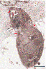
Toxoplasma Gondii is a protozoan that commonly occurs in many types of environments (Harker et al., 2013). Toxoplasma Gondii could be both extra-cellular and intracellular (see figure one). However, studies have shown that it is predominantly intracellular (Feustel, Meissner, & Liesenfeld, 2012). A patient’s immune system is always critical in making sure there is a low risk of pathogenic infections from Toxoplasma Gondii because, unlike other pathogenic subspecies, such as T. b. gambiense and T. b. rhodesiense, Toxoplasma Gondii could be dormant within a patient’s system for a long time (Feustel et al., 2012). However, an immunity compromise is likely to activate it. When it attacks, it could have a high prevalence among the infected population (Masocha & Kristensson, 2012). For example, studies in Europe and Africa have shown that the parasite could have a prevalence of up to 90% (Stanford University, 2015). Comparatively, studies based in Australia and Japan show that its prevalence could be exceptionally low (Stanford University, 2015). Infections by Toxoplasma Gondii usually show no known symptoms, but patients who have compromised immunity could suffer some of its worst effects (Berenreiterova, Flegr, Kubena, & Nemec, 2011). This group of patients also suffers a high risk of infection to the central nervous system (CNS). Patients who have congenital disorders are also likely to suffer a high risk of CNS infection (Carey, Westwood, Mitchison, & Ward, 2004). Encephalitis and other neurological diseases are common effects of advanced stages of CNS infection (Stanford University, 2015). Deleterious effects are also synonymous with CNS infections (Carey et al., 2004). Another common cause of infection is the immunosuppressing effect of HIV and AIDS. This condition usually reactivates latent infections.
Mechanism of Penetration through the Blood-brain Barrier
When Toxoplasma Gondii penetrates the blood-brain barrier, it usually causes cysts (Stanford University, 2015). However, before this process occurs, the protozoon usually invades its host through absorption via the small intestine (Coombes et al., 2013). Thereafter, it affects macrophages to make sure that the host’s immune system is incapacitated. The macrophage host cell is usually instrumental in preventing Toxoplasma Gondii infection because it helps in killing the protozoa through an oxidative burst, occasioned by nitric oxide production (Stanford University, 2015). However, Toxoplasma Gondii prevents this process from occurring by infecting the macrophages (Masocha & Kristensson, 2012). Nonetheless, infected monocytes are immune from this action because they could still produce nitric oxides and kill the parasite intracellularly (Stanford University, 2015). Lymphokines are also immune to the efficient macrophage-killing mechanism of Toxoplasma Gondii. This way, they could activate macrophages and other cells to attack Toxoplasma Gondii (Stanford University, 2015). Toxoplasma Gondii also risks elimination by the immune system through its invasive intuition mechanism, through the small intestine, which could damage the intestinal epithelium, thereby causing an inflammatory response. This outcome could easily trigger a full-blown immune response from the host’s body (Coombes et al., 2013). To minimize the possibility of elimination, Toxoplasma Gondii has to develop better biological responses that would not trigger the immune system, or prevent its elimination by protecting itself from a full-blown immunological response. Stanford University (2015) says the presence of gram-negative bacteria is the main trigger of the full-blown immunological response. Lipopolysaccharide production usually characterizes this response (Coombes et al., 2013). Toxoplasma Gondii prevents its elimination by possessing a gene that suppresses lipopolysaccharide production (Zhou et al, 2005). Cytokine production is usually the product of such a process. Such defensive mechanisms usually allow Toxoplasma Gondii to thrive by reproducing and penetrating the blood-brain barrier (Coombes et al., 2013).
Despite the above explanations regarding how Naegleria Fowleri penetrates the blood-brain barrier, researchers do not fully understand the mechanisms used by Toxoplasma Gondii to penetrate the blood-brain barrier (Stanford University, 2015; Siddiqui & Khan, 2014). However, preliminary reports show that the protozoa use two primary methods – attachment to white blood cells and attachment to endothelial cells (Carey et al., 2004). By attaching itself to white blood cells, Toxoplasma Gondii could penetrate the blood-brain barrier because white blood cells have a free passage through this barrier (Stanford University, 2015). However, this process is difficult for the Toxoplasma Gondii to accomplish because the presence of macrophages could complicate its attempt. Experts say the infiltration of the blood-brain barrier through the endothelial cells contributes to the highest cases of Toxoplasma Gondii brain infections, compared to infiltration through the white blood cells (Coombes et al., 2013). However, the host’s innate and acquired immune responses are likely to suppress attempts to penetrate the blood-brain barrier (Stanford University, 2015). To counter this response, Toxoplasma Gondii produces tachyzoites that form immune-resistant pseudocysts (Carey et al., 2004). These cysts usually assume a dormant nature until reactivation occurs. Usually, the host’s innate immune system prevents these cysts from activating. Indeed, the medical evidence shows that these cysts attempt to rapture and multiply, but the host’s immune system prevents them from doing so (Masocha & Kristensson, 2012). Usually, this process happens under normal conditions, but experts still do not know the mechanism used by the host’s immune system to prevent this action (Lachenmaier, Deli, Meissner, & Liesenfeld, 2011).
People who have suppressed immunities often suffer the highest risks of cyst reactivation (Masocha & Kristensson, 2012). This failure may allow a full-blown infection to occur. The lack of antibodies in the brain often worsens this situation because CNS infections are likely to persist (compared to when the infection would occur in a different organ or part of the body) (Stanford University, 2015). Based on these dynamics, acute infections among patients who suffer from suppressed immunities are likely to occur in the central nervous system (Lachenmaier et al., 2011). Such infections are likely to cause severe pathogenesis, which draws a strong link with taxoplasmosis (Stanford University, 2015). Relative to this observation, Stanford University (2015) says, “Toxoplasmosis in the brain often consists of necrotizing encephalitis and associated inflammation, to which microglia respond by forming nodules in attempts to contain the infection” (p. 20). Often, suppressed immunities compromise this process. When such a situation occurs, Toxoplasma Gondii replicates faster than the host’s immunity could control it (Stanford University, 2015). The clinical effects of such compromised immunity are usually severe in affected patients.
Mechanism of Taenia Solium
Taenia Soliumn is the main causative agent for epilepsy. It usually affects the central nervous system through the spread of intermediate larvae from the porcine tapeworm, Taenia Solium (Motarjemi, 2013). Unlike other parasites highlighted in this paper, there is sufficient evidence explaining how Taenia Solium penetrates the blood-brain barrier. However, before delving into the details surrounding its penetrative mechanism, it is pertinent to understand that most people get infected by Taenia Solium by eating contaminated food (usually pig products) (Velázquez-Moctezuma et al., 2014). Taenia solium usually penetrates the blood-brain barrier by entering the blood system through the small intestine (Sun et al., 2014). Although infections are asymptomatic, infected people become hosts of Taenia Soliumn cells that continue to multiply in their systems. When multiplication occurs and Taenia Solium proportions increase in the host’s circulatory system, they affect the central nervous system and the subcutaneous tissue (Singh & Prabhakar, 2002). The Taenia Soliumn lives in the patient’s immune system as cysts that could stretch between 10 to 20 millimeters (Quinones-Hinojosa, 2012). To evade the host’s immune system, the parasite usually modulates it.
Taenia Soliumn could penetrate the blood-brain barrier because it covers its cysts with host-derived molecules. At the same time, it usually secretes immunomodulatory enzymes to counter the effects of the host’s immune system. These penetrative mechanisms usually occur by passing through the lymphatic system and bloodstream. This penetrative mechanism differs from that of Toxoplasma Gondii and Naegleria Fowleri as discussed below.
Discussion
Although few parasites, or pathogens, could penetrate the blood-brain barrier, evidence from this paper shows that Toxoplasma Gondii, Naegleria Fowleri, and Taenia Solium employ some immunosuppressing techniques to minimize the effect of the host’s immune system. Furthermore, some of these pathogens have preventive mechanisms for stopping antibodies from attacking them. Their strength is further elevated when hosts are suffering from immunosuppressive conditions, such as HIV/AIDS, or when they are taking immunosuppressing drugs (Singh & Prabhakar, 2002). Although the mechanisms used by the pathogens to infiltrate the blood-brain barrier may vary, their target is the same – the central nervous system. This target is preferable because immunity (in this system) is low (based on the possibility of an inflammatory response) (Stanford University, 2015). However, minimized immunity does not mean that the pathogens do not have to manage the host’s immunity because they do. Based on this limitation, penetrating the blood-brain barrier is often a common challenge for these microscopic brain eaters. For those that can do so, evading the host’s immune system, or suppressing it, are usually common characteristics of their penetrative mechanism (Estanol, Juarez, Irigoyen, Gonzalez-Barranco, & Corona, 1989). Nonetheless, the mechanisms used by the three species studied are not common. For example, this paper has shown that these species use different invasive mechanisms to attack their hosts. Toxoplasma Gondii and Taenia Solium enter the host’s blood supply system through the small intestine. Comparatively, Naegleria Fowleri enters the host’s blood supply system through the host’s nasal cavity. Indeed, by attaching to the upper wall of the host’s nasal cavity, the pathogens gain access to the frontal lobes. This paper has also shown that these pathogens use different mechanisms to penetrate the blood-brain barrier. For example, it has been shown that Toxoplasma Gondii penetrates the blood-brain barrier by attaching to white blood cells and the host’s endothelial cells. Comparatively, Naegleria Fowleri penetrates its host’s blood-brain barrier through the olfactory epithelium and the cribriform plate. Lastly, Taenia Soliumn penetrates the host’s blood-brain barrier by covering its cysts with host-derived molecules. This assessment alone shows that the three pathogens do not use the same mechanisms to penetrate the blood-brain barrier (see table one). Another area showing the differences in mechanisms used by the three pathogens is how they manage the host’s immune systems. Indeed, evidence from this paper shows that these pathogens endure the immune attack from the host’s system by selectively using different characteristics of the host’s immune system for their posterity. This way, the parasites penetrate the blood-brain barrier usually at the expense of their hosts. For example, Taenia Soliumn protects itself from the host’s immune system by modulating it. Comparatively, Toxoplasma Gondii kills the macrophage host cell, which would have otherwise created antibodies to fight the pathogen. Furthermore, this pathogen evades the host’s immune system by disguising itself as one of the host’s cells. Comparatively, Naegleria Fowleri copes with the host’s immune system by damaging inhibitor cells. These different mechanisms show that the three pathogens use different mechanisms for penetrating the blood-brain barrier and staying there (see table 2). The only area of commonality among the three pathogens is their target (brain tissue). They attack it by penetrating the blood-brain barrier and infecting the central nervous system. Findings from this paper also show that although the host’s immune system may struggle to contain infections in the brain tissue, the pathogens could replicate themselves much faster than the immune system could kill them (Sun et al., 2014). These mechanisms are adaptive methods used by pathogens to damage the host’s brain cells (Sun et al., 2014). Immunosuppression creates the greatest vulnerability to such pathogens. Therefore, the risk of pathogenic infection across the blood-brain barrier remains low (for healthy people) (Paredes et al., 2013). Comprehensively, although the findings presented in this paper are factual, it is important to note that they are not comprehensive because they stem from existing literature, which has not fully explained the mechanisms used by the above-mentioned pathogens to penetrate the blood-brain barrier.
Comprehensively, the mechanisms for penetration, outlined in this paper could be instrumental in expanding medical knowledge about the mechanisms used by these parasites to invade the blood-brain barrier. Consequently, they could inform valuable medical interventions about the same. Similarly, this research could be useful in developing highly efficacious treatment methods. This process is the first step in developing preventive treatment methods for stopping these parasites from causing further harm to their host’s central nervous system. However, there is a need to undertake more research to fine-tune the results depicted in this study.
Tables and Figures


Table one: Differences in Invasive mechanisms for species
Table 2: Differences in coping mechanism for Species
References
Berenreiterova, M., Flegr, J., Kubena, A., & Nemec, P. (2011). The Distribution of Toxoplasma gondii Cysts in the Brain of a Mouse with Latent Toxoplasmosis: Implications for the Behavioral Manipulation Hypothesis. PLoS ONE, 6(12), 1-14.
Burri, D. C., Gottstein, B., Zumkehr, B., Hemphill, A., Schürch, N., Wittwer, M., & Müller, N. 2012. Development of a high-versus low-pathogenicity model of the free-living amoeba Naegleria fowleri. Microbiology, 158, 2652-2660.
Cardona, A., Restrepo, B., Jaramillo, J., & Teale, J. (1999). Development of an Animal Model for Neurocysticercosis: Immune Response in the Central Nervous System Is Characterized by a Predominance of gd T Cells. J Immunol, 162(1), 995-1002.
Carey, K. L., Westwood, N. J., Mitchison, T. J., & Ward, G. E. (2004). A small-molecule approach to studying invasive mechanisms of Toxoplasma gondii. Proceedings of the National Academy of Sciences of the United States of America, 101, 7433–7438.
Chauhan, A., Sun, Y., Pani, B., Quenumzangbe, F., Sharma, J., Singh, B. B., & Mishra, B. B. 2014. Helminth Induced Suppression of Macrophage Activation Is Correlated with Inhibition of Calcium Channel Activity. PloS one, 9, 101-123.
Chávez-Munguía, B., Villatoro, L., Omaña-Molina, M., Rodríguez-Monroy, M., Segovia-Gamboa, N., & Martínez-Palomo, A. (2014). Naegleria fowleri: Contact-dependent secretion of electrondense granules (EDG). Experimental parasitology, 142, 1-6.
Coombes, J. L., Charsar, B. A., Han, S.J., Halkias, J., Chan, S. W., Koshy, A. A.,…Robey, E. A. (2013). Motile invaded neutrophils in the small intestine of Toxoplasma gondii-infected mice reveal a potential mechanism for parasite spread. Proceedings of the National Academy of Sciences of the United States of America, 110, 1913–1922.
Davson, H. (1993). An Introduction to the Blood-brain Barrier. New York, NY: CRC Press.
Estanol, B., Juarez, H., Irigoyen, I., Gonzalez-Barranco, D., & Corona, T. (1989). Humoral immune response in patients with cerebral parenchymal cysticercosis treated with praziquantel. J Neurol Neurosurg Psychiatry, 52(1): 254-257.
Feustel, S., Meissner, M., & Liesenfeld, O. (2012). Toxoplasma gondii and the blood-brain barrier. Virulence, 3(2), 182–192.
Harker, K., Ueno, N., Wang, T., Bonhomme, C., Liu, W., & Lodoen, M. (2013). Toxoplasma gondii modulates the dynamics of human monocyte adhesion to vascular endothelium under fluidic shear stress. Journal of Leukocyte Biology, 93(5), 789-800.
Khan, N. (2008).Acanthamoeba and the blood–brain barrier: the breakthrough. J Med Microbiol, 57(9), 1051-1057.
Lachenmaier S., Deli, M., Meissner, M., & Liesenfeld, O. (2011). Intracellular transport of Toxoplasma gondii through the blood-brain barrier. J Neuroimmunol, 232(1), 119-30.
Manno. R. (2012). Emergency Management in Neurocritical Care. London, UK: John Wiley & Sons.
Masocha, W., & Kristensson, K. (2012). Passage of parasites across the blood-brain barrier. Virulence, 3(2), 202–212.
Motarjemi, Y. (2013). Encyclopedia of Food Safety. New York, NY: Academic Press.
Pardridge, W. (2006). Introduction to the Blood-Brain Barrier: Methodology, Biology and Pathology. Cambridge, UK: Cambridge University Press.
Paredes, A., Campos, T., Miguel, L., Rivera, M., Dorny, P., Mahanty, S.,…Cass, Q. (2013). In Vitro Analysis of Albendazole Sulfoxide Enantiomers Shows that (_)-(R)-Albendazole Sulfoxide Is the Active Enantiomer against Taenia solium. Antimicrobial Agents and Chemotherapy, 57(2), 944-949.
Quinones-Hinojosa, A. (2012). Schmidek and Sweet: Operative Neurosurgical Techniques: Indications, Methods and Results (Expert Consult – Online and Print). Philadelphia, PA: Elsevier Health Sciences.
Roy, S., Metzger, R., Chen, J., Laham, F., Martin, F., Kipper, S.,… Visvesvara, G. (2014). Risk for Transmission of Naegleria fowleri From Solid Organ Transplantation. American Journal of Transplantation, 14(1), 163–171.
Siddiqui, R., & Khan, N. (2014). Primary Amoebic Meningoencephalitis Caused by Naegleria fowleri: An Old Enemy Presenting New Challenges. PLoS Negl Trop Dis, 8(8): 1-8.
Singh, G., & Prabhakar, S. (2002). Taenia Solium Cysticercosis: From Basic to Clinical Science. New York, NY: CABI.
Stanford University. (2015). How CNS-Invasive Parasites Evade the Brain’s Immune System. Web.
Sun, Y., Chauhan, A., Sukumaran, P., Sharma, J., Singh, B. B., & Mishra, B. B. 2014. Inhibition of store-operated calcium entry in microglia by helminth factors: implications for immune suppression in neurocysticercosis. Journal of neuroinflammation, 11, 210-212.
Toney, D., & Marciano-Cabra, F. (1994). Membrane Vesiculation of Naegleria fowleri Amoebae as a Mechanism for Resisting Complement Damage. Journal of Immunology, 152, 2952-2959.
Velázquez-Moctezuma, J., Domínguez-Salazar, E., & Gómez-González, B. (2014). Beyond the borders: The gates and fences of Neuroimmune interaction. New York, NY: Frontiers E-books.
Zhou, X. W., Kafsack, B. F., Cole, R. N., Beckett, P., Shen, R. F., & Carruthers, V. B. (2005). The opportunistic pathogen Toxoplasma gondii deploys a diverse legion of invasion and survival proteins. Journal of Biological Chemistry, 280, 34233-34244.