Introduction
The procedure adopted in the study was a standard way of assessing neurological functions of the nervous system. It is one of the methods that are used in hospital settings to estimate the state of nerves in the body. Andreassi (2000) argues that an essential component in nerve conduction is the speed at which impulses and responses are transmitted along the motor or sensory pathways.
The nervous system is an important part of the body because it is involved in responding to stimuli in the external environment (Misulis 1997). The CNS is important in mediating stimuli processing in the brain and spinal cord. On the other hand, the PNS is characterised by thousands of nerves that are appear as long fibres. They are used in linking various parts of the CNS. It has been shown that the long fibres form motor neurons, which are involved in coordinating voluntary movement (Misulis 1997). The PNS also contains nervous components that mediate involuntary functions. In healthy individuals, the transmission of impulses should be within standard ranges of speeds. Thus, a nerve conduction velocity is the pace of conducting electrochemical impulses along neurons. A number of factors could influence the speed of nerve conduction. For example, disease, age and sex, among other factors could make different persons have diverse velocities of conducting impulses (Izhikevich 2000). It has been shown that changes to the constant velocity of nerve conduction could be an indication of damage (Andreassi 2000).
The architecture of the nervous system shows that both motor and sensory axons are found in the same neurons. Thus, if a disease affects either of the axons, then the other type of axon could also be affected. Patients who present with nerve diseases could be characterised by various symptoms. For example, they could feel numbness when some stimuli are applied to their bodies. Also, they may have tingling thoughts even in the absence of stimulus. Individuals whose nerves are damaged could have altered responses with regard to temperature and pain. In fact, they could be exposed to elevated temperature and a lot of pain, but they show little responses. Nerve damages have been shown to result in loss of sense with regard to position and vibration. The nervous system may be damaged and appear through acute or chronic manifestations.
Demyelination is a common cause of diseases in the nervous system (Misulis 1997). The process involves a loss of the myeln sheath that surrounds axons. Due to the importance of myelin sheath in increasing the speed of nerve conduction, loss of the component results in reduced speed of conducting impulses along neuron fibres (Kandel, Schwartz & Jessell 2000). Nerve diseases that affect axons are caused due to the interruption of conduction velocity with regard to demyelination. In fact, deterioration of the destruction of the myelin sheath affects axons that are involved in conducting impulses at similar speeds. Thus, the initial functions that are compromised in nerves following nerve diseases are action potentials that have similar speeds of transmission. Some of these are tendon reflexes and vibratory sensation (Kandel et al 2000; Misulis 1997). The reduction of nerve conduction velocity was investigated in the experiment.
Procedures
All procedures in the experiment were carried out keenly while recording all readings. The recording was important to ensure that all details were captured and used in the required calculations.
Sensory nerve conduction
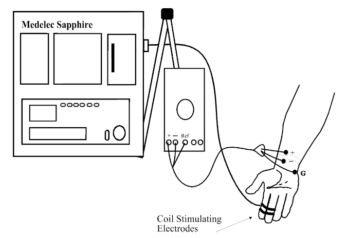
Ulnar and median nerves were located and the skin surface was prepared by cleaning it with alcohol. Coil stimulating electrodes were attached to recording electrodes and ground electrode. The skin impedance was tested using a metre to ensure that the readings were within the recommended range before starting the nerve conduction velocity measurement procedure. The equipment set by averaging various parameters in order to obtain the right measurements. The subject was made to seat comfortably while stretching the right arm and relaxing to minimise the chances of introducing muscle artefact during measurements. The equipment was set to start stimulating the subject while recording observations. Various intensities of stimuli were applied to the subject with the aim of recording different response readings. In addition, different stimuli were used to obtain the threshold of stimulus that could elicit the best response. Nerve action potential readings were recorded. The next step involved measuring the distance between the stimulating and recording electrodes. In addition, the latency of the action potential responses were recorded. From the readings, the conduction velocities of the motor fibres were calculated.
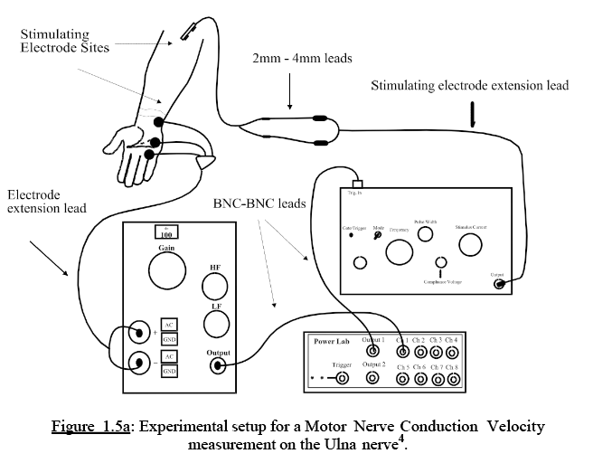
The ulnar nerve was located on the subject taking part in the experiment. A preparation with regard to the recording and ground electrodes was made by abrading the skin and cleaning it using alcohol. In addition, a daub of conductive gel was applied on the skin surface to facilitate the sensitisation of the skin. The stimulating electrodes were soaked in saline solution. The skin impedance was tested using the impedance metre. The isolated stimulator in the equipment was used to determine the threshold of the subject. It was important to make the order of stimulating electrodes and recording electrodes the same. In order to ensure that the stimulating electrodes were kept wet, they were dipped in saline solution. The recording electrodes were placed on the flashy palm using the proper orientation of the negative and positive electrodes of the equipment. The stimulating electrode was placed on the nerve sight behind the elbow. At that point, it was essential apply different stimuli from the equipment. With regard to the experiment, a stimulus intensity of applied on about 5muA. The intensity of current that was used to stimulate the subject was increased. At the same time, the recording electrode around the skin region to determine the best stimulation site.
Results and discussion
This section of the report includes the results of the experiments and key discussion points.


Figure 3. A table that summarises the calculations of nerve conduction velocities using distances between peaks and latency.
The velocities were calculated using the following steps:
Velocity = distance/latency
MNCV = (d2-d1)/((t2-t1)
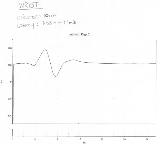
For the wrist, velocity = (10-0)/(7.58-3.77) = 2.62467 m/s.
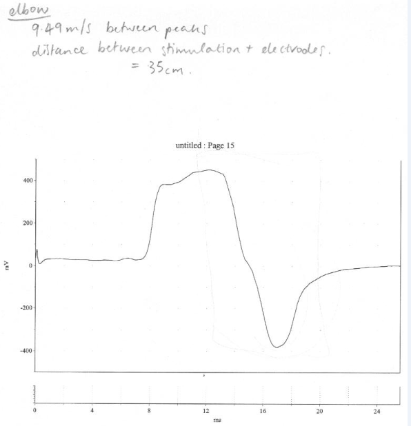
For the elbow, velocity = (35)/(9.49) = 3.688 m/s


The results obtained in the experiment show important findings with regard to the nerve conduction velocity and latency values. The latency value obtained from the wrist experiment falls within the normal range that is found in healthy persons. However, the nerve conduction velocity of 2.62 m/s is below the standard value that is documented in physiology literature. A number of reasons could cause variations. First, it could be due to experimental errors that could have occurred during the experiment. Errors result in wrong findings that cannot be used to make conclusions about clinical observations. For example, wrong readings could have been taken. Doing calculations with wrong readings increases the chances of obtaining wrong results.
Second, there could have been wrong targeting of the site with the best stimulation responses. The effect of a wrong region could be that a region that was identified could not give the best readings of responses. This could have been avoided during the preparation stage when the skin of the subject was being prepared for measurement.
Third, there could be a possibility the conduction gel applied on the skin did not help in conducting stimuli onto the skin of the subject. Thus, it could have acted as a barrier to effective sensitisation of the skin.
Fourth, it would also be important to consider the variation noted the results from a pathological perspective. There could be nerve damage in the subject used in the experiment. Although the subject might not be aware of the pathological state of the nerves, the damage might be in the early stages of chronic damage. Myelin sheath of the axons of the neurons could be characterised by some form of degeneration (Misulis 1997). Damage of the myelin sheath interferes with the normal transmission of impulses along axons. This is due to the disruption of the patterns of the nodes of ranvier that act as crucial points of facilitating the speed to impulse transmission. Thus, the pace at which the impulses that were induced in the subject were transmitted was relatively slow and could not fall below the normal range.
Conclusion
The experiment showed that latency values could be normal in a subject whose nerve conduction velocity values are abnormal. The findings indicated that it is essential to conduct experiments that are free from errors. Based on the results, it could be concluded that the subject was suffering from some of nerve damage or errors were common in the procedures.
References
Andreassi, JL, 2000, Psychophysiology: Human behavior and physiological response. Psychology Press, London, United Kingdom.
Izhikevich, E M, 2000, ‘Neural excitability, spiking and bursting’, International Journal of Bifurcation and Chaos, vol. 10, no. 06 pp. 1171-1266.
Kandel, ER., Schwartz, JH, & Jessell, TM, Eds, 2000, Principles of neural science, McGraw-Hill, New York, NY.
Misulis, KE, 1997, Essentials of clinical neurophysiology , 2nd ed. Butterworth-Heinemann , Upper Saddle River, NJ.