Introduction
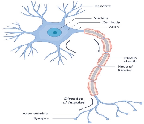
The nervous system is the major communicating, regulatory, controlling system in the body. The nervous system is the center of all mental activities including memory, learning, and thought. In conjunction with the endocrine system, the nervous system is in charge of maintaining and regulating homeostasis.
The objective of this laboratory report is to dissect, identify the internal cavity of a rat, and examine the different sensory organs of a rat. Additionally, this report examines the neuron anatomy from the prepared microscope slides in the laboratory. Although humans and rats seem to be entirely different, they tend to share some physiognomies, which may include biological, psychological, or behavioral. Like other body systems, the nervous system comprises organs such as the ganglia, nerves, spinal cord, and brain.
Background Information
According to Snyder and others (2018), the nervous systems are developed from glia and neurons. Neurons are the key functional cells while glia plays a range of primary support roles. Subsequently, the nervous system develops as an interconnected network of cells. The brains of vertebrates are the largest nervous organs, while other simple animals such as jellyfish have nerve nets (Mastorakos & McGavern, 2019). Researchers posit that self-awareness or consciousness is one critical concept of the nervous system, and helps to differentiate between perception and sensation. This report explores the anatomy and physiology of the nervous system of a rat.
According to the National Institute of Neurological Disorders and Stroke (2018), the nervous systems are different in every animal and are crucial for interpreting and detecting information, regulating body functions, making important decisions, and movement. Therefore, the physiology and anatomy of the nervous systems of rats and that of humans are considerably similar. For that matter, rats tend to sense and react similarly to their stimuli, such as touch, injury, or disease.
Through testing rats, healthcare personnel has made tremendous steps in the advancement of medicine (MedlinePlus, 2016). Through dissecting a rat we can see exactly how similar a rat’s anatomy and physiology of the nervous system is to that of a human being (National Institute of Neurological Disorders and Stroke, 2018). This report reviews the cellular structures and functions and investigates several fundamental neural networks within the body. Furthermore, this report uses a combination of exercises and models, physiological, anatomical dissection, and microscopic analysis to examine the nervous system.
Relevant Observation
This experiment involves two exercises, which include: (1) Dissecting a rat to examine the internal organs including the sensory organs, and (2) examining the microscope slides to learn about the anatomy of neurons using a light microscope. Several relevant observations were made from this experiment. From the first exercise, the major sensory organs were all observed including the eyes, skin, tongue, nose, ears, the spinal cord, the brain stem, and the brain. From the second exercise, the following parts were observed; myo-neural junction, cerebellum, and the spinal cord.
Hypotheses
- The physiology and anatomy of the nervous systems of rats and that of humans are considerably similar.
- Rats sense and react similarly to their stimuli, such as touch, injury, or disease.
Materials and Methods
Equipment and Supplies
- Forceps
- Scissors
- A dissecting tray
- Disposable gloves
- Probes
- Dissecting pins and twins
- Freshly killed rat
- Arm bone cutter
- Microscope
- Masks
Steps
Exercise 1
- Pin the rat to the wax of the dissecting tray by placing the dorsal side down
- By using scissors make an incision on the median line of the animal
- Complete incision from pelvic to the jaw
- Try to expose the underlying muscles
- By using scissors cut through the abdominal skin
- Now lift the skin and cut through the muscle layer from the pubic region to the bottom of the rib cage
- Near rib cage make two lateral cuts so that the thin membrane inside should be visible
- The membrane is the diaphragm which separates thoracic and abdominal cavities
- Cut the diaphragm where it attaches to the ventral ribs
- Lift the ribs to see the thoracic cavity clearly
- Cut across the flap at the level of the neck and remove the rib cage
- To expose the brain and the roots of the cranial nerves make a hole at the junction of frontals and parietals with a pointed arm bone cutter
- Remove the bones carefully not to damage the roots of the 12 pairs of cranial nerves
Exercise 2
Steps
- Turn the revolving nosepiece to click the lowest power objective lens into position.
- Put the microscope slide on the stage and hold it using the stage clips.
- Observe the objective lens and the stage sideways and adjust the focus knob to move the stage up. Move it upwards to maximum but the objective lens should not touch the coverslip.
- Look through the eyepiece and adjust the focus knob to focus the image.
- Tune the condenser for maximum light intensity.
- Adjust the microscope slide until the specimen is in the field of view.
- Use the coarse and fine adjustment knobs to focus the specimen and readjust the light intensity and condenser.
- When the image comes into clear view with the lower power objective, you can readjust to the next objective lenses, either the specimen or lighting can be adjusted. Repeat steps 3 to 5 with the higher power objective lens if the image is not clear.
- Lower the stage when finished, click the lower power lens into position, and remove the slide (MedlinePlus, 2016).
Results
Exercise 1
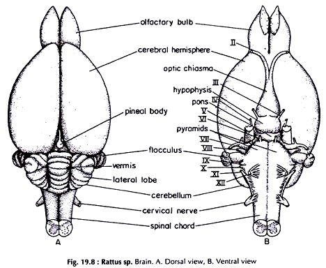
The major sensory organs observed include the eyes, skin, tongue, nose, ears, the spinal cord, the brain stem, and the brain. These organs have specialized cells and tissues which receive raw stimuli and translate them into signals that can be used by the nervous system (NINDS, 2018). Please see the figures below:
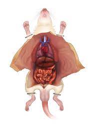
Similar to other body systems, the nervous system comprises organs such as the nerves, spinal cord, and brain. Consecutively, these organs comprise tissues such as connective tissues, blood, and nerves. Collectively, these tissues and organs perform the complex activities of the nervous system.
Exercise 2
The microscope slides showed several details of the neuron anatomy. The neuron anatomy of a rat and that of a human are closely similar in several ways including:
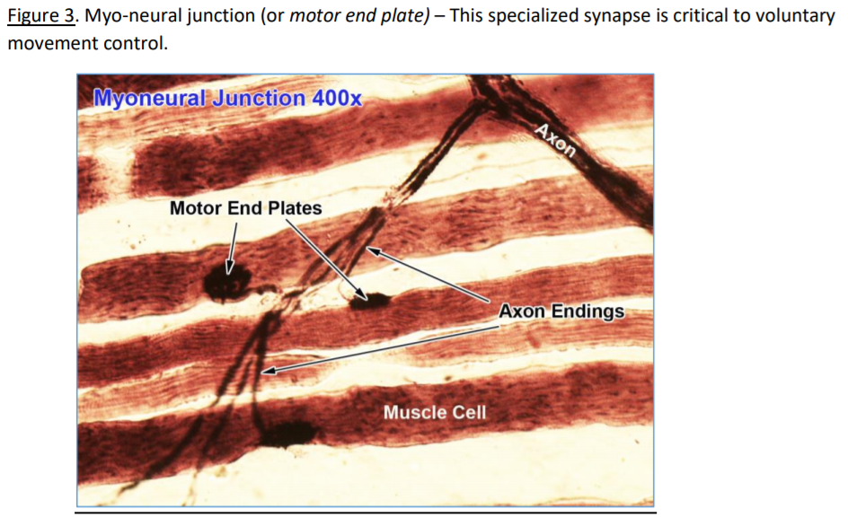
- Motor end plate –when viewed through the microscope, the synaptic connection between a skeletal muscle cell and motor neuron can be seen.
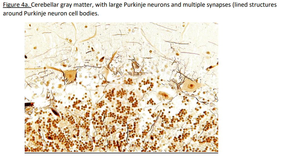
- Cerebellum – in both cases of the human and rat’s nervous systems, the cerebellum has folded gray and white matter that can be distinguished with the eyes.
- Spinal cord – in both cases, a cross-sectional view of the spinal cord shows central gray matter and peripheral gray matter.
Data Presentation and Analysis
The data are in this report are presented in form of figures and pictures. According to the figure and pictures shown above of the nervous systems of a rat, it can be seen that a rat’s nervous system is closely related to that of a human being (Zeisel et al., 2018). It is determined that there are similarities in the shapes of the organs and the systems and the types of the organs responsible for the nervous system functions. It was examined that the nervous system of a rat and a human being are closely related.
Discussion and Conclusion
From the first exercise, the major sensory organs were all observed including the eyes, skin, tongue, nose, ears, the spinal cord, the brain stem, and the brain. According to Snyder and others (2018), these organs have specialized cells and tissues which receive raw stimuli and translate them into signals that can be used by the nervous system.
The nervous system has two main parts: the Central Nervous System (CNS) and the Peripheral Nervous System (PNS). The CNS is comprised of the spinal cord and the brain while the PNS comprises nerves branching from the spinal cord and extend to all parts of the body. The nervous system transmits information from the brain to the rest of the body and vice versa (Mastorakos & McGavern, 2019). As a result, the activities of the nervous system control the ability to think, see, breathe, and move.
From the second exercise, the following parts were observed; myo-neural junction, cerebellum, and the spinal cord. Firstly, the Motor end plate can be viewed at the synaptic connection between a skeletal muscle cell and motor neuron (NINDS, 2018). Additionally, structures like motor endplates, myelin sheath of Schwann cells on axons, skeletal muscle cells with sarcomere, and axons; all look similar in both the human nervous system and in the rat nervous system.
Secondly, the Cerebellum has folded gray and white matter that can be distinguished with the eyes (MedlinePlus, 2016). Subsequently, within the gray matter, there are three distinct layers of neurons namely: molecular, Purkinje, and granular layer, which discloses organizational structures. Lastly, a cross-sectional view of the spinal cord shows central gray matter and peripheral gray matter, which in turn has small central canal with ependymal glia.
Zeisel et al. (2018) state that large motor neuron cells are situated in the ventral gray matter. Moreover, sensory neurons are located in the dorsal root ganglia, and their axons develop into the roots of the spinal nerves. The nervous system keeps us in touch with the internal and external environment (temperature, sound, light, pH, and oxygen levels). Therefore, just like in humans, the rat’s nervous system performs the same functions- sensory, integrated, and motor functions.
References
Mastorakos, P., & McGavern, D. (2019). The anatomy and immunology of vasculature in the central nervous system. Science immunology, 4(37).
MedlinePlus. (2016). Neurosciences. Web.
National Institute of Neurological Disorders and Stroke. (2018). Brain basics: Know your brain. Web.
Snyder, J. M., Hagan, C. E., Bolon, B., & Keene, C. D. (2018). Nervous system. In Comparative anatomy and histology (pp. 403-444). Academic Press.
Zeisel, A., Hochgerner, H., Lönnerberg, P., Johnsson, A., Memic, F., Van Der Zwan, J., Haring, M., Braun, E., Borm, L.E., Manno, G.L., Codeluppi, S., Furlan, A., Lee, K., Skene, N., Harris, K.D., Hjerling-Leffler, J., Arenas, E., Ernfors, P., Marklund, U., & Linnarsson, S. (2018). Molecular architecture of the mouse nervous system. Cell, 174(4), 999-1014.