The evolution of the sun
The sun, which is the center of the solar system, is the main source of light and energy for the entire earth (Merali & Skinner, 2009). The sun has been described as a huge ball of fire and all the eight planets of the earth revolve around it. The ball of fire is made up of several elements, which include hydrogen, helium, iron, nickel, oxygen, silicon, and sulfur among others.
The sun has a temperature of approximately 5,780 K and is approximately 1.496×10¹¹ m from the earth (Merali & Skinner, 2009). This paper will discuss the sun’s formation and its patterns of movement.
It is difficult to tell the exact period of formation of the sun but it was approximated to have been between ten and twenty thousand million years ago (Merali & Skinner, 2009). The Nebular theory also called the Condensation theory is one of the theories that explain the origin and formation of the sun. Astronauts suggest that the hydrogen present in the sun was influenced by the big bang theory.
This justifies the assumption that the sun came into being around the same period as the rest of the universe (Merali & Skinner, 2009). According to the big bang theory, hydrogen gas through condensation formed huge clouds. These clouds after contracting they formed the present galaxies.
The role of motion and bodies involved in the formation of the sun
However, some hydrogen gas did not contract and was floating freely in our galaxy. Due to some incidents, the free hydrogen gas also contracted leading to the formation of the sun and the entire solar system. It is said that the sun and the solar system later turned into a slowly spinning molecular cloud (Merali & Skinner, 2009).
This cloud was composed of hydrogen, helium molecules, and dust (Merali & Skinner, 2009). Gravity caused the cloud to compress a process called Helmholtz contraction (Merali & Skinner, 2009). The inability of gases to balance against self-gravity was the major cause of the contraction and this is what astronauts refer to as jeans instability (Noguchi, 1999).
As the compression process went on the speed of rotation also increased and the high-speed spinning caused the cloud to flatten forming a giant disk-like shape (Noguchi, 1999). Astronauts believe that the majority of the Sun’s mass collected at its center, which led to the formation of a gas sphere (Noguchi, 1999).
The sphere further influenced compression by attracting materials from the disc a factor that led to an increase in the temperatures and pressure from within the sphere (Noguchi, 1999).
The collusion of these particles turned the kinetic energy into heat. This caused the nebular to accumulate great heat at its center where most of its mass was. This led to the collapse.
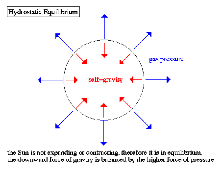
However, the process could have continued but hydrostatic equilibrium prevented it. As the heat increased at the center of the cloud, the pressure rose and influenced an outward net force (Noguchi, 1999). Below is a diagram showing the hydrostatic equilibrium (Noguchi, 1999)

The sun’s motion
The sun is the closest star to earth compared to all other stars of our galaxy. Every day, the sun rises in the east and reaches its maximum height when it crosses the meridian at noon (Merali & Skinner, 2009). It takes the sun approximately 24 hours to move from the noon position to the noon position in the next day (Merali & Skinner, 2009).
In that case, the noon position is whenever the sun crosses the meridian in a day (Merali & Skinner, 2009). The position of the horizon is not constant throughout the year but changes from time to time (Merali & Skinner, 2009)
The sun moves along the ecliptic completing a full 360 degrees in a year, which is approximately 365.24 days (Noguchi, 1999). The path that the sun follows through the stars is known as the ecliptic (Noguchi, 1999). The ecliptic and the celestial equator both traverse at two points. These two points are the vernal equinox and the autumn equinox (Merali & Skinner, 2009).
Around 21st march each year, the earth crosses the celestial equator going northwards at the vernal equinox ‘spring’ (Merali & Skinner, 2009). It later crosses the celestial equator going southwards at the autumnal equinox around 22 September each year (Noguchi, 1999).
During this time when the sun is in the celestial equator equinoxes, the entire world experiences equal days and equal nights for the two days (Merali & Skinner, 2009). During the season of spring and summer, the sun is usually above the celestial, which makes the earth experience more than 12 hours of daylight (Noguchi, 1999). This is because the sun does not rise at the exact east hence making a longer arc north of the celestial equator.
On the other hand, when the earth is below the celestial equator the earth experiences shorter hours of daylight. This is the season of autumn and winter and is caused by the sun rising in the southeastern making a shorter arc south of the celestial equator (Merali & Skinner, 2009). Nonetheless, the path of the sun depends highly on the date and the latitude the observer is at (Merali & Skinner, 2009).
Copernicus’s, Kepler’s, Galileo’s, and Newton’s inventions
Copernicus was the first to explain the movement of the sun. In his theory, he indicated that the earth rotates daily around its axis and revolves around the sun for a period of about 365 days (Noguchi, 1999). Isaac Newton’s law of gravity was a great input in explaining how the sun could curve inward into the elliptical paths.
Through Nicolaus Copernicus findings, the heavenly bodies do not share the same center since the earth rotates around the sun and the moon around the earth (Merali & Skinner, 2009). He argues against the belief that the sun rotates around the earth.
Johannes Kepler’s second law suggests that the speed of a planet increases when it gets closer to the sun (Merali & Skinner, 2009). The farther a planet is from the sun therefore the slower its speed. He also came up with a law that suggested that the orbits of the planets were not circular but elliptical in shape (Merali & Skinner, 2009). Galileo Galilei confirmed Copernicus theory to be true by affirming the rotation of the rotation of the sun around the earth.
Conclusion
This essay has extensively discussed and given scientific theories, that supports the formation of the sun and its movement. The paper explains the sun’s formation and its properties. The motion of the sun causes seasons that have been clearly outlined in this research.
References
Merali, Z., & Skinner, B. J. (2009). Visualizing Earth Science. Hoboken, NJ: Wiley.
Noguchi, M. (1999). Early Evolution of Disk Galaxies: Formation of Bulges in Clumpy Young Galactic Disks. Astrophysical Journal, 514 (1), 77–95.