Introduction
Ovarian cancer is a disease that starts in and affects the female reproductive organs, the ovaries. It is a very dangerous disease and common cancer among women that claims a lot of lives as compared to other kinds of cancer that affect the female reproductive system. This is especially due to the fact that in its early stages, it either shows minimal nonspecific signs or none at all making it difficult for its early diagnosis and intervention which worsens the condition (Buist et al., 1994).
This report gives an in-depth discussion of ovarian cancer including a description of its pathology, the methods used in its diagnosis especially the imaging methods, how the pathology looks like on images, the effects of the pathology on procedures, and finally its treatment and prognosis.
Description of Pathology
Pathology entails a study of disease including everything that pertains to it for instance the causes, its development path, symptoms, and the effects, what affects its spread or curing process among other manifestations of the disease. It is also termed pathobiology. Here is the pathology of ovarian cancer. Although the effect of ovarian cancer especially in terms of the deaths caused is well known, the exact cause is not known.
However, several factors have been deemed to contribute to the risk of developing ovarian cancer, for instance, the lesser children a woman has and the later in life she bears children, the higher the chances of developing ovarian cancer. Bearing children reduces the risk of developing ovarian cancer. BRCA1 and BRCA2 genes are also responsible for causing this disease though they contribute a small percentage.
An individual’s or family history may also boost ovarian cancer occurrences. Age is also an issue of concern as ovarian cancer tends to be suffered much by older women who are 55 years of age and above as opposed to the rate in the younger women. Its existence in women below the age of 40 years is minimal. Pills administered for birth control also reduce the risk of developing ovarian cancer (National Comprehensive Cancer Network, 2009).
According to Fayed (2010), there are common known factors that tend to increase the risk of ovarian cancer. It is for example a disease that is common in women who have reached menopause, have a family history of either breast cancer, ovarian cancer, or colon cancer, or those who had suffered from breast cancer sometimes back, have not borne any children, suffer from obesity, have undergone a therapy that entails estrogen replacement, have used some fertility drugs for long periods among other factors. Although ovarian cancer originates from the ovary, it affects other parts of the body as it spreads out for instance to the pelvic region, the intestines, and the lower part of the back.
The symptoms of ovarian cancer are usually hard to be identified during the first stages of the disease and even when they appear they are not specific. They are only notable at the advanced stages of the disease when its effects have caused a lot of damage to the body. This usually makes it hard to recover from the ordeal as compared to when it could be detected in its initial stages.
Some of the signs associated with ovarian cancer include abdominal discomfort, high rates of bloating, pain in the reproductive system especially around the pelvis, low appetite, virginal bleeding, unexpected weight loss, and gains, excessive fatigue is caused by the disease’s tendency of draining the body’s energy, pains, and discomfort during sexual intercourse, pressure, and pains in body parts for instance the stomach, legs, backaches, and irregular menses among others (Morch, 2009).
Diagnostic Methods
There exist numerous methods that could be applied in the diagnosis of ovarian cancer. The initial steps that a doctor may take in the event of suspecting the existence of ovarian cancer include some pelvic examination, CA-125 test, and ultrasound. Physical examination, CT scan, a transvaginal ultrasound, magnetic resonance imaging (MRI), biopsy, and laparotomy could also be applied.
Surgery could also be part of diagnosis apart from being a treatment option. Magnetic resonance imaging (MRI) is a medical test that helps in the diagnosis and treatment of various diseases ovarian cancer is one of them. It produces pictures of various body structures through the use of a strong magnetic field displayed on a computer. The images are then scrutinized and a conclusion is made from the observation.
It is a very effective method and allows for the identification of the presence of certain diseases that could otherwise not be detected using other imaging methods for instance computed tomography (CT) and X-ray. Although MRI has various benefits for instance the fact that it does not entail any exposure to ionizing radiation, it also has some risks like allergic reactions and nephrogenic systemic fibrosis especially in patients with kidney problems. Some medical devices in a patient’s body may interfere with the effectiveness of the MRI procedure (Anonymous 2010).
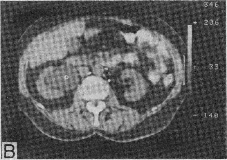
Computed tomography (CT) or CAT scanning is an imaging method that can produce images of bone, blood vessels, and tissues at a go. It works on the same principle that is applied on X-rays but instead of the images being presented on a film, a detector which is in a banana shape is used to present and measure the profile. The physician then makes conclusions based on the images in regard to the presence of ovarian cancer and the extent to which it has spread. CT has the following advantages over other imaging methods; it is relatively cheap, it is relatively fast, and widely available.
Treatment and Prognosis
Prognosis entails the prediction of the possible course and outcome of a particular disease including the chances of recovery or survival that exist. The option to be chosen in the treatment of ovarian cancer is highly dependent on the extent to which cancer has spread which is gauged by the stage and grade of cancer. Treatment of the disease mostly involves surgical treatment and also through the use of chemicals. As another method of treatment, medical doctors expose the affected area to radiation.
Ovarian cancer is a disease that is treatable especially in the event that it is detected early enough hence avoiding further damages. Surgery works by removing the tumors and growth that could have been caused by ovarian cancer. Some organs of the body could also be removed to avoid further infections which may cause more complications. The surgery also entails some relocation of body parts for instance the pancreas and the liver. Surgery too enhances the success of chemotherapy through the removal of the body parts.
Chemotherapy, on the other hand, works by preventing the further spread of the disease. This is done through administering some drugs for instance carboplatin, paclitaxel, that suppress cancer through elimination or killing of the cancer cells that could have survived the surgery process. It is done either prior to all after the surgery but the latter is more common. The frequency and duration of the chemotherapy depend on an individual patient for instance in regard to the health status like resistance or reaction to some medications or even the stage of the disease in the individual.
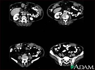
Radiation therapy is rarely used in the treatment of ovarian cancer and in most cases it is applied with the main aim of reducing the symptoms associated with the disease (Hayat, 2009). Some of the images that show the conditions of ovarian cancer are shown below.

Peritoneal and ovarian cancer, CT scan: this shows a CT scan series of the lower abdomen showing ovarian cancer that has spread to the peritoneum.

Conclusion
Ovarian cancer is a critical disease that affects a large percentage of women worldwide. The problem is that there are no explicit measures that can be taken to prevent its occurrence. All in all, there are some factors that could be considered by women in an effort of reducing the chances of suffering from the disease. Some of them can however not apply to all women and cannot just be done for the major aim of reducing the risk. They include bearing children as research shows that giving birth even though it is to one child reduces the risk of ovarian cancer, use of oral contraceptives, and tubal ligation among other measures.
Reference List
Anonymous, (2010). MRI of the Body (Chest, Abdomen, Pelvis). New York: Radiology Society of North America.
Buist, R.M et al. (1994). Comparative Evaluation of Diagnostic Methods in Ovarian Carcinoma with Emphasis on CT and MRI. Gynecologic Oncology Volume 52, Issue 2, pp 191-198.
Fayed, L. (2010). The Causes, Symptoms, Treatment and Prevention of Ovarian Cancer. Web.
Hayat, A.M. (2009). Methods of Cancer Diagnosis, Therapy, and Prognosis: Ovarian Cancer, Renal Cancer, Urogenitary Tract Cancer, Urinary Bladder Cancer, Cervical Uterine Cancer, Skin Cancer, Leukemia, Multiple Myeloma and Sarcoma. USA: Springer.
Morch LS, Lokkegaard E, Andreasen, AH, Krüger-Kjaer S, & Lidegaard, O. (2009). Hormone Therapy and Ovarian Cancer. JAMA, 302:298-305.
National Comprehensive Cancer Network (2009). National Comprehensive Cancer Network Clinical Practice Guidelines in Oncology: Ovarian Cancer, vol. 2.