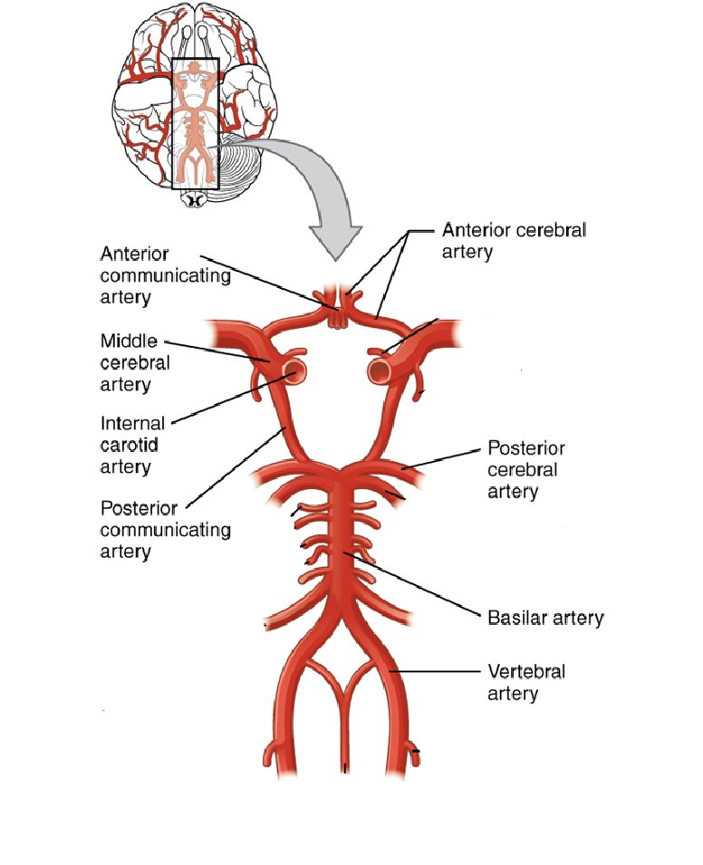
The diagram above is an occipital view of how blood is supplied to the brain. The central nervous system (CNS) needs continuous micro-circulation with oxygen and sustenance. The highest part of the CNS with an oxygen demand is the brain. The ischemic cells in the brain die from lacking oxygen. The vertebral and internal carotid arteries are the two paired arteries that deliver blood to the brain. These arteries start in the neck and go up to the skull. The terminal branches of these arteries create an anastomotic circle within the cerebral vault known as the Circle of Willis. Branches emerge from this circle, supplying the bulk of the cerebrum. Smaller branches from the vertebral arteries supply other areas of the CNS, such as the pons and spinal cord.
An anterior communicating artery crowns the circle of Willis and acts as a link-up between the two anterior cerebral arteries. The posterior communicating artery is a limb of the internal Carotid artery that connects it to the posterior cerebral artery. Both the anterior and posterior arteries are termed “Linking receptacles.” The internal carotid artery emerges where the sinistral and dextral common carotid arteries bifurcate at the cavernous sinus. Its main purpose is to supply blood rich in oxygen to the significant anatomy: of the brain and eyes, which is made possible through the emergence of the ophthalmic artery and anterior choroidal arteries. This artery then bifurcates into the middle and anterior cerebral arteries. The motion and sensuous palliums of the upper limb and face are predominantly supplied by the middle cerebral arteries, providing Broca’s area in the prepotent frontal lobe and Wernicke’s in the prepotent temporal lobe. The anterior cerebral arteries, on the other hand, dispense oxygenated blood to the brain portion that is principally behind the motor and sensory control of the lower limbs.
The basilar artery emerges from the junction of the vertebral arteries at the bottom of the skull, where the head converges with the neck. It makes sure that oxygen and nutrients reach critical areas such as the cerebellum, brainstem, and occipital lobes. The brainstem aids in reflex actions such as respiration, eupepsia, and sleep patterns; the cerebellum, on the other hand, sees that the body has balance, pose, integration, and articulation. The Vertebral artery is responsible for transporting blood from the brain to the spinal cord. Their emergence is at the subclavian arteries, underneath the clavicle; the right subclavian finds its base at the brachiocephalic artery, while the left subclavian is directly at the aorta. These vertebral arteries traverse independently in the sinistral and dextral regions of the vertebral column in the neck.
All the arteries in this diagram are significant in that they contribute to the well-being of the cerebral-spinal structures; however, the basilar artery proves to be the most significant in the organism. For the body to be balanced in movements and posture, there must be enough supply in the brain to transmit neurons to the spine, which is responsible for the support of the body as a whole. Clinically, the disruption of blood oxygenation in the brain leads to stroke of the spine and the cerebellum, which affects the body, disabling both reflex and voluntary actions. The contributing factors to this lack of supply of blood to the brain are stroke embolism, thrombosis, hypoperfusion, and excessive bleeding.