Aims and objectives
This report is written with regard to two laboratory practicals. The two laboratory practicals aimed at confirming whether or not patients’ serum samples were characterized by antibodies that could react against the gastric proton pump. The techniques that were mainly used were gel electrophoresis, Western blotting and immune-histochemistry tests. The experiments also focused on assessing the category of patients who had pernicious anemia.
Introduction
Pernicious anemia is a disease known as the last presentation of chronic gastritis, which is an autoimmune disease that negatively affects the stomach (Toh, van Driel & Gleeson1997). The disease is characterized by inflammation of the stomach and over secretion of autoantibodies (Oliva-Hemker, Berkenblit, Anhalt & Yardley 2006; Toh et al 1997). The autoantibodies could be an intrinsic factor and proton pump. These proteins are secreted by the parietal cells of the stomach. Diminishing of the mass of the stomach (gastric atrophy) is the commonest symptom that characterizes patients suffering from pernicious anemia (Annibale et al 2000; Oliva-Hemker et al 2006). The physiological cause of the disease has been correlated with the activity of autoantibodies that negatively impact intrinsic factors, which is essential for the absorption of vitamin B12 (Toh et al 1997). If the activity of the intrinsic factor is compromised, then the amount of vitamin B12 absorbed is greatly reduced. Hydrochloric acid is an important agent in the initial stages of digestion. Research shows that the secretion of the acid into the stomach is mediated by H+/K+-ATPase. H+/K+-ATPase is a proton pump that has been shown to be involved in essential transport pathways within cells. Chemical studies with regard to the structure of the enzyme have shown that it has unique beta and alpha subunits (Toh et al 1997). The subunits have been implicated in reactions with autoantibodies that are found in the serum of human beings (Toh et al 1997).
Methods
SDS-PAGE and Western blotting
The study of components of protein mixtures is conducted through the use of SDS-PAGE. When proteins are being studied, they are dissolved in sodium dudecyl-sulfate (SDS), which binds to the portions of proteins that are hydrophobic. The purpose of SDS is to make the proteins linear and achieve a negative charge. Disulfide bonds that hold chains of peptides are broken through the action of reducing chemicals such as 2-beta mercaptoethanol. When such proteins are run on gel electrophoresis, they migrate from the negative side of the equipment to towards the positive portion, where they could combine with the opposite charges at the anode. Western blotting is carried out to determine the identity of proteins. Essentially, Western blotting involves the transfer of proteins separated through the SDS-PAGE onto the membrane. The commonly used membranes are made of nitrocellulose.
PAGE
A premade polyacrylamide gel was placed in an electrophoresis tank, that contained a running buffer. So that a stomach protein sample isolated from the stomach of a mouse could be prepared, 50µl of 5x SDS buffer was added to a tube that contained 200 µl of stomach protein. The tube contents were thoroughly mixed to obtain a homogenous solution. A molecular weight marker was used so that it could give references to the distances traveled by protein samples. In fact, the marker was added to the first well of the gel. Each well was loaded with 25 µl of the protein samples. The electrophoresis was run for 1 hour. Afterward, the proteins that were separated through the electrophoresis process were transferred onto a nitrocellulose membrane. Once the transfer was completed, the membrane was placed into a vessel that contained 50mL 0.1% Ponceau stain and placed on the rocker for 1 minute. The membrane was labeled using a pencil so that the correct interpretations regarding the source of proteins could be made. Next, the membrane was rinsed using 50mL 0.1M NaOH. This was done to clear the stain on the nitrocellulose membrane. It was also rinsed in 50mL TBS. Nonspecific binding of proteins was prevented from occurring by the addition of 50mL TBS. The TBS was mixed with about 5% skim milk powder. After labeling the membrane, it was stored so that it could be used in the experiments that could be conducted later.
Assessment of the morphology of the stomach of a mouse
Rehydration was done in running tap water for about 30 minutes. Afterward, the slide containing the rehydrated cells was incubated in hematoxylin solution for 2 minutes, rinsed in water and shortly dipped in alcohol that contained acid. To avoid the contents of the slide from being exposed to the prolonged effects of the acid, the slide was immediately rinsed in water. Afterward, the slide was washed in eosin Y for 4 minutes and in 90% ethanol for 30 seconds. Consequently, it was washed in absolute ethanol for 30 seconds. The slide was prepared for viewing under the microscope so that the morphology of the stomach cells could be established. Proper labeling of diagrams with regard to the images obtained was adopted. It was important to maintain proper labeling so that contamination could be reduced significantly.
Continuation of Western blotting
The nitrocellulose membrane that contained the separated proteins was retrieved from the storage pouch. About 100 µl of the sera was added onto each strip of the membrane and the membrane portions were incubated at room temperature for 1 hour. Later, the strips were washed in TBST buffer and rocked. They were placed onto a parafilm. About 100 µl of the secondary antibody was distributed onto the films. The strips were washed in TBS and rocked. The membrane was placed into a Chemidoc apparatus that could aid the detection of the substrate. The equipment yielded two images for each test sample.
Immune-peroxidase staining of mouse stomach sections
The presence of particular proteins (antigens) in the tissue section was determined through the immuno-histochemistry technique. The method is commonly applied in detecting antibodies in serum samples. In the practical, the presence of anti-proton pump antibodies was being determined using the serum. The sections of the mouse stomach contained the proton pump antigen. Once unbound serum was washed away, the section was incubated with a secondary antibody that was very specific for human immunoglobulin molecules. The secondary antibody was conjugated to protein (an enzyme), which could convert the substrate to a colored product that could be easily recognized through the reading apparatus.
The following procedures were used in this part of the practical: The slide with the mouse sections was placed in a container with xylene for 2 minutes and in ethanol for 2 minutes. In order to remove the washing reagents, the slide was rinsed in tap water. The endogenous peroxidase activity that could negatively impact the quality of the results was removed by washing the slide in 0.3% hydrogen peroxide and PBS for 30 minutes. Afterward, the slide was washed for 6 minutes in PBS. Sheep anti-human (50 µl) anti-immunoglobulin solution was added to the section and incubated for 45 minutes. The slide was washed in PBS for 6 minutes. The next step involved the addition of 100 µl of Dab substrate to the section and incubation was done for 10 minutes. In order to remove the washing buffers, the slide was washed by dipping into hematoxylin for only 3 seconds and in water for 30 seconds. The slide was washed in ethanol for 2 minutes. A small drop of DPX mounting medium was placed on the section and covered with a coverslip. However, the excess mounting medium was cleared from the slide through the application of clean cotton material. The prepared slide was viewed under the microscope and the images were recorded.
Results
SDS-PAGE
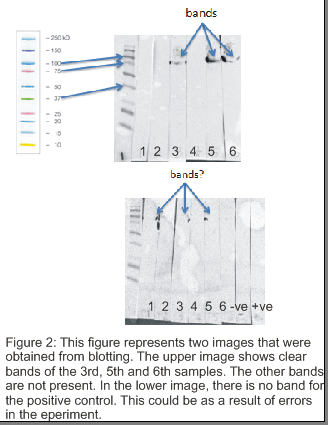
Figure 1 shows the results that were produced from PAGWE. The results show the different migration distances covered by the molecular marker, negative and positive controls, and the samples being tested. The expected size of the protein elements that were being identified by the sera from pernicious anemia patients was about 90-100kD. Thus, samples 3, 5 and 6 exhibited that they contained the protein components, which were characterized by bands within that range (90 to 100kD). The results that were obtained with regard to SDS-PAGE are shown in figure 1. The size of the protein is commonly associated with the alpha subunit of the proton pump, which is found in gastric cells that are important in the absorption of vitamin B12 in human beings. ELISA test could be used as an alternative to test for the presence and amounts of serum proteins in samples. ELISA would detect the presence of the proteins on the premises of antigen-antibody reactions.
Immunochemistry
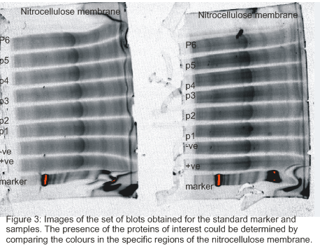
Results that were obtained from immunoblotting are presented in figure 2 and figure 3. The upper image in figure 2 gives the values of the anticipated size of the proteins that were being tested. It also shows the samples that contained the proteins of interest. Particularly, samples 3, 5 and 6 had the proteins that were being assessed. In fact, the images of the bands with regard to the three positive samples were very clear. The image on the lower side of the figure indicates that the positive control did not have any band (product formed). This could have been caused by an error in the laboratory. The sheep anti-human Ig-HRP conjugate that was used in the Western and immuno-histochemistry was specific to the proton pump antigen. A transgenic could be the most appropriate animal in which the antibodies were raised. The animal was antigenic to allow a high degree of purity of the antibodies. The antibody was raised in a transgenic mouse to increase its specificity for the antigen. The enzyme that was conjugated in the immunoblotting technique was horseradish peroxidase, which was used so that it could change the substrate to the product with a specific color.


Immune-peroxidase staining of mouse stomach sections
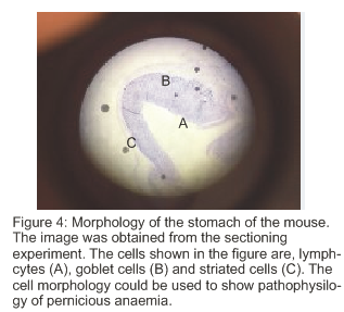
The results that were obtained from the sectioning of the stomach of the mouse are presented in figure 4. The figure shows a cross-section of the stomach that could be assessed to learn about the effects of pernicious anemia. Different cells are indicated in the figure. The morphology of the stomach is important in the progression of the disease.

Discussion
The results show that the proton pump is an important factor in the diagnosis of pernicious anemia. Vitamin B12 insufficiency has been demonstrated in the disease reported in this report. All the aims of the practicals were achieved. Thus, the findings could be used to provide a better understanding with regard to pernicious anemia. However, there were some shortcomings in the practicals, which negatively impacted the quality of the findings. For example, the positive control used in Western blotting did not produce any bands. This raises the issue of the authenticity of the results. In addition, most of the samples produced bands that were not proper bands on the basis of molecular sizes. One of the reasons for the observation could be improper antibody incubation. Also, the problem could have resulted from contamination and poor labeling of samples and other materials in the laboratory. The experiment could be improved in the future by ensuring that proper antibody incubation approaches would be adopted (Howanitz & Howanitz 2001). In addition, proper labeling and decontamination strategies would be utilized to improve the quality of results.
References
Annibale, B, Lahner, E, Bordi, C, Martino, G., Caruana, P, Grossi, C,… & Delle Fave, G, 2000, ‘Role of Helicobacter pylori infection in pernicious anemia’, Digestive and Liver Disease, vol. 32, no. (9), pp. 756-762.
Howanitz, JH, & Howanitz, PJ, 2001, ‘Laboratory results in timeliness as a quality attribute and strategy’, American journal of clinical pathology, vol. 116, no. (3), pp. 311-315.
Oliva-Hemker, M, Berkenblit, GV., Anhalt, GJ, & Yardley, JH, 2006, ‘Pernicious anemia and a widespread absence of gastrointestinal endocrine cells in a patient with autoimmune polyglandular syndrome type I and malabsorption’, Journal of Clinical Endocrinology & Metabolism, vol. 91, no. 8, pp. 2833-2838.
Toh, BH, van Driel, IR, & Gleeson, PA, 1997, ‘Pernicious anemia’, New England Journal of Medicine, vol. 337, no. 20, pp. 1441-1448.