Introduction
This paper is a review of the historic anatomy of the pancreas. The pancreas is one of the most important organs in the digestive system. The paper seeks to analyze the development and discovery of the pancreatic functions in the body. The paper seeks to elaborate clearly the anatomy and structure of the pancreas and the specialized functions it performs in the body.
The report seeks to identify the functions of other organs that help in the digestive process where the pancreas is dominantly involved. This paper seeks to unveil the historic discovery of the pancreas and to document the structural and functional aspect of the organ.
Anatomy

The pancreas was one of the last organs that were discovered in the research conducted to explain the digestion process. The development of the pancreas research continued from 200 AD until the final details where established in 1664. The functions of the pancreas were examined using the pancreatic fistula of dogs. Nonetheless, the digestive functions of the pancreas were not discovered until 1844.
Anatomists discover that the pancreas was excreting a juice that emulsifies fats and digests starch. During this period, a demonstration to show the digestive action of the pancreas juice on sugar, fats, and proteins was presented. This experiment was carried out using the pancreas fistula of a dog. Although in 1876 the pancreatic functions were believed to be driven by an enzyme, this was later disapproved after the discovery of secretin in the juice.
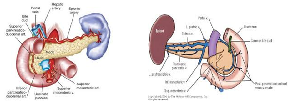
After researchers discovered secretin that was contained in the pancreatic juice, they disapproved the belief of enzymatic action. Its body is elongated towards the left within the aorta and attached on it by the peritoneum of the lesser sac. Its head is positioned on the right and it is placed at one end where the duodenum bends. The pancreas is divided into three parts, a neck, body and a tail. The tail stretches all the way to the gastric face of the spleen. Here, the body of the pancreas stretches leftward on the outer side of the aorta. It is retroperitoneal and it is held against the aorta by the peritoneum of the lesser sac.
The pancreas is positioned at the back of the abdomen and other organs including the liver, the small intestines to mention, but a few engulf it. The pancreas is shaped like a flat pear but its two ends are not equal in size. The bigger side, which is normally on the right hand side, is called the head while the smaller end is the tail. Several blood vessels surround the pancreas. They include the superior mesenteric artery, the superior mesenteric vein, the portal vein, and the celiac axis.
Structure
The body of the pancreas and its tail connect without a clearly protruding point of contact between them. One of the observable characteristics of the tail is its mobility as its tip stretches out to the spleen. The tail at this point is contained and held between the splenorenal ligaments by the spleen, artery, and vein.
The function of the splenocolic ligaments is to attach the splenic mobile characteristic of the colon to the spleen. This function brings the colon closer to the pancreas where its digestive functions begin to take place. The pancreas has very vital function on a number of abdominal structures.
Through the formation of unit by the bile duct, the duodenum, and the pancreas, digestion is enhanced in the stomach. The bile duct can be found on the right hand side of the gastroduodenal artery in the inner walls of the stomach. All these veins supply blood not only to the pancreas but also to other vital abdominal organs in the body.
The excretion of exocrine glands is vital in the digestion process. The glands are the driving force that allows smooth and effective digestion. They are found in the pancreas. During digestion, food enters through the mouth and goes directly to the stomach. Here, the pancreatic juices are released into the systems of ducts that lead to the pancreatic duct.
Variation of the pancreas
The size of the pancreas is not constant in all mammals. It differs depending on the embryological development of one individual to another. The pancreas develops in the form of two buds embroiled on the duodenum. The ventral bud is supposed to rotate fully under normal circumstances but in some cases, it may not complete a full cycle. This may lead to a situation called pancreatic divism.
Histological structure
The pancreas is made up of tissues that contain endocrine and exocrine functions, and this separation is observable when the pancreas is placed under a microscope. The tissues that contain endocrine functions are observed under the microscope as lightly stained groups of cells, and their scientific name is islets of Langerhans. The darker stains seen under the microscope form another group of cells that are called Acini. These groups of cells are set in lobes at odds by an emaciated tough wall.
The secretory cells of each group of tissues surround a small-intercalated canal. To perform their functions effectively, these cells are made up of many small granules of zymogens can be seen. A solitary sheet of columnar cells shapes the ducts. Since they are large, several layers of columnar cells can be clearly seen.
Ductal structure
Near the tail of the pancreas, a duct is formed from other ductile that helps in drainage. These ducts are responsible for draining the lobules of the glands. The pancreas has accessesory ducts that help it to communicate with the main duct. These ducts are located in the bile duct and in the minor papilla, which is situated in close proximity to the second duodenum. This close proximity allows efficient communication during digestion within the duodenum. The pancreas plays a major role in this process.
Conclusion
This research has reviewed clearly and efficiently the discovery of the pancreas. The paper has started by outlining the history and foundation of pancreatic discovery from the 200 AD. The development and experiments that led to the discovery of the pancreas and its functions have been chronologically presented.
The paper further outlines the anatomy of the pancreas in a detailed manner to explain its function in the digestion process. Several other organs that are related to the pancreas and its functions have also been discussed including the duodenum, the intestines, and the bile duct. This paper has explicitly discussed the histological structure of the pancreas in detail. The ductal structure of the pancreas has also been studied and outlined in this comprehensive research work.
References
Anatomy and Histology of the Pancreas. Jpck. 2013; 1(1): 1-8. Web.