Introduction and Patient’s History
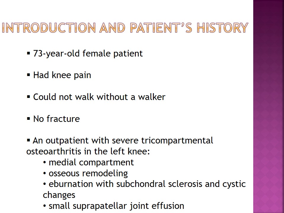
- 73-year-old female patient.
- Had knee pain.
- Could not walk without a walker.
- No fracture.
- An outpatient with severe tricompartmental osteoarthritis in the left knee:
- medial compartment;
- osseous remodeling;
- eburnation with subchondral sclerosis and cystic changes;
- small suprapatellar joint effusion.
In this case, a patient was a 73-year-old female with knee pain as her chief complaint. She could not walk without a walker and had to be transformed to the table with the help of a wheelchair. Although no recent fractures were reported, the radiologist’s reading presented enough information about the patient’s condition, including severe tricompartmental osteoarthritis in her left knee (medial compartment is more pronounced compared to other parts). Additional health problems were osseous remodeling, eburnation with subchondral sclerosis, some cystic changes, suprapatellar joint effusion, and dystrophic calcification.

Pathology
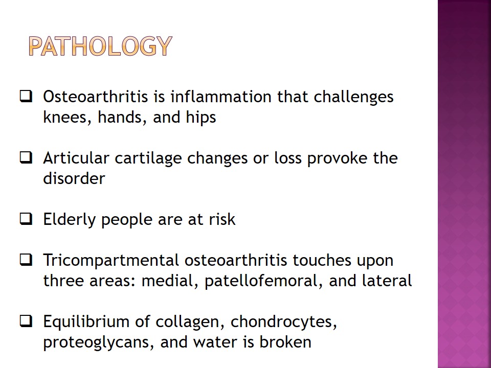
- Osteoarthritis is inflammation that challenges knees, hands, and hips.
- Articular cartilage changes or loss provoke the disorder.
- Elderly people are at risk.
- Tricompartmental osteoarthritis touches upon three areas: medial, patellofemoral, and lateral.
- Equilibrium of collagen, chondrocytes, proteoglycans, and water is broken.
Osteoarthritis is a common form of arthritis that may affect many people from different parts of the work. This type of inflammation damages joints and is frequently observed in knees, hands, or hips. Knee osteoarthritis happens because of wear and tear of articular cartilage or even its progressive loss (Hsu & Siwiec, 2019). Elderly women and men are usually the populations at risk. Tricompartmental osteoarthritis is characterized by changes in three compartments at once: medial (inside the knee), patellofemoral (across the kneecap), and lateral (outside the knee). There are several important components in articular cartilage, including collagen, chondrocytes, proteoglycans, and water, and when its equilibrium cannot be maintained, inflammation is developed, causing pain and bone changes (Hsu & Siwiec, 2019). Traumas, aging, weight, and genetics are the major causes of this disorder.

Imaging Techniques and Treatment
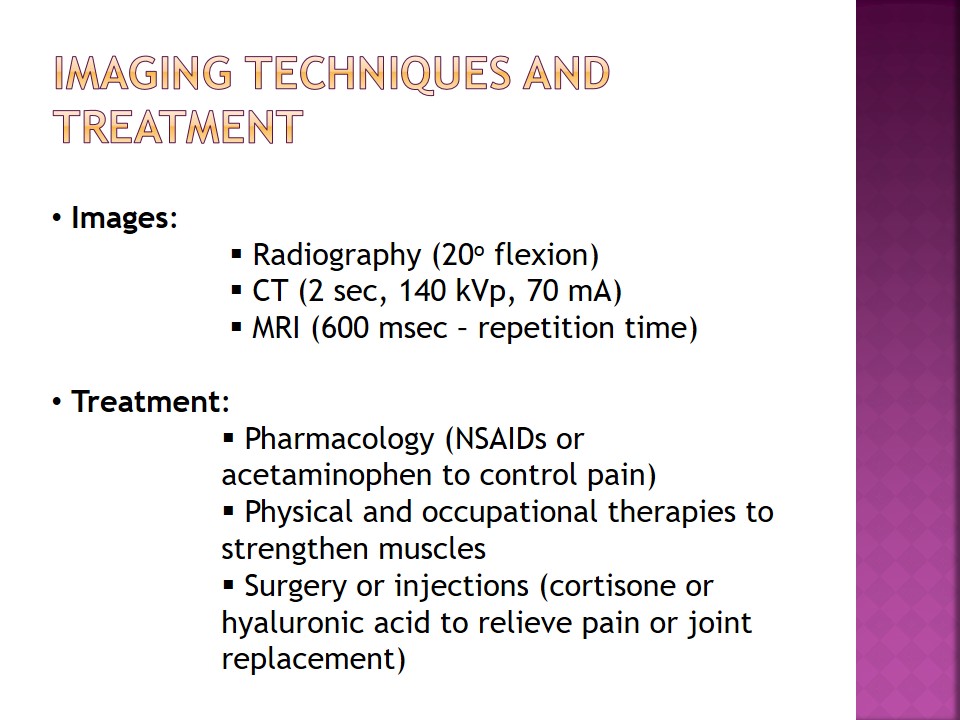
- Images:
- Radiography (20o flexion);
- CT (2 sec, 140 kVp, 70 mA);
- MRI (600 msec – repetition time).
- Treatment:
- Pharmacology (NSAIDs or acetaminophen to control pain);
- Physical and occupational therapies to strengthen muscles;
- Surgery or injections (cortisone or hyaluronic acid to relieve pain or joint replacement).
The identification of the disorder is possible with the help of radiographs, computed tomography, or magnetic resonance imaging. MRI is the most comprehensive assessment tool to identify and classify a disorder, as well as promote tissue contrast (Li, Amano, Link, & Ma, 2016). As soon as the diagnosis is given, a treatment plan should be developed. In this case, the patient has to consider pharmacological treatment, including NSAIDs or acetaminophens like Tylenol to control pain, or physical and occupational therapies to strengthen muscles, modify activities, and consider weight changes. Finally, surgery to replace joints or injections with cortisone or hyaluronic acid to relieve pain is recommended.

References
Hsu, H., & Siwiec, R. M. (2019). Knee osteoarthritis. Web.
Li, Q., Amano, K., Link, T. M., Ma, C. B. (2016). Advanced imaging in osteoarthritis. Sports Health, 8(5), 418-428.