Introduction
Due to its prevalence and associated poor health outcomes, anemia turns out to be a serious public health concern. According to Nagao and Hirokawa (2017), anemia is defined as a condition when a person does not have enough red blood cells (RBCs) in the body, which results in a lowered ability to carry oxygen. As a rule, people with anemia feel weakness and fatigue that is hard to control. There are several types of anemia, and each of them has its causes and clinical manifestations.
Microcytic anemia occurs when the number and the size of RBCs are smaller than normal (iron deficiency), and macrocytic anemia happens when these cells are larger than normal (alcoholism). Normocytic anemia is characterized by normally sized RBCs that are low in number (kidney problems). Therefore, it is important to study this condition and its forms to predict complications and treat patients effectively. Anemia is not a diagnosis but a syndrome that makes millions of people visit doctors, and an understanding of its types contributes to its prevention.
Morphologic Classification
Several ways can be offered to classify anemia, its causes, and outcomes. Being observed in routine blood cell enumeration, anemia’s classification is based on such red cell morphology parameters as the number of RBCs and the mechanism of causation (Chaparro & Suchdey, 2019). The distinctions between macrocytic, microcytic, and normocytic anemias depend on the results of a diagnostic approach where mean corpuscular volume (MCV) and mean corpuscular hemoglobin (MCH) is considered (Braunstein, 2019). As has already been mentioned, there are three main types of anemia, however, the presence or absence of some vital changes and biological factors introduces new subgroups with specific manifestations.
Macrocytic Anemia
When vitamin B12 or folate deficiency is observed, doctors say that a person may have macrocytic anemia (or macrocytosis). In adults, this condition is defined when an RBC number is more than 100 femtoliter (fL) (Nagao & Hirokawa, 2017). Depending on different morphologic characteristics, macrocytic anemia could have additional names. For example, normochromic macrocytic anemia means that the MCV stays within the normal limits, and the MCH is decreased.
Pernicious anemia is determined by vitamin B12 deficiency when absorption is impossible. This condition is rare and was known as a deadly disease because no effective treatment was available. Nowadays, the replacement of vitamin B12 in the body is an available treatment option. Finally, macrocytic anemia is also known as folate (vitamin B-9) deficiency when people do not have access to folate-rich products, face increased requirements (pregnancy or lactation), or take medications (phenytoin) (Nagao & Hirokawa, 2017). The development of RBC is extremely important for the human body, and the choice of a diet remains a critical factor in the required nutrient absorption.
Microcytic Anemia
Compared to macrocytic anemia, the size of RBCs in macrocytic anemia is smaller than normal. The MCV in patients with microcytosis is lower than 80fL (Adamu et al., 2017). In the case of hypochromic anemia, both the RBC and MCH levels are decreased. Pre-menopausal women are at high risk of this condition because they lost a considerable amount of blood regularly, which reduces the presence of iron in the body.
The causes of this type of anemia vary, and one of the most common is iron deficiency (Chaparro & Suchdey, 2019). The bone marrow needs iron stores to support growth and development during such periods as infancy or pregnancy. Iron deficiency occurs when an individual is not able to recognize the source of bleeding (acute blood loss) or because of chronic slow blood and iron loss (menstruation).
Sideroblastic anemias are characterized by abnormalities in iron metabolism due to failed erythropoiesis. This condition may be hereditary when the RBC mitochondria cannot support heme synthesis or acquired because of alcohol or drug intake. Thalassemias introduce a group of conditions (alpha and beta) with defects in “the synthesis of one or more of the globin chains” – α-globin and β-globin chains, respectfully (Chaparro & Suchdey, 2019, p. 27). The deletion of at least one gene in chromosome 16 (alpha) or 11 (beta) causes a decrease in hemoglobin, which results in the microcytic anemia.
Normocytic Anemia
There are also cases when an individual has normal-sized RBCs, but their number is low. In this situation, a doctor says that normocytic anemia is observed as a particular type of anemia. As well as other types, this condition may be inborn or acquired. Aplastic anemia is rare and caused by vital stem cell damage in the bone marrow. Post-hemorrhagic occurs when a person suddenly losses blood, which causes the inability to control the level of hemoglobin and acute metabolism complications.
Hemolytic anemia is a condition during which the destruction of RBCs is faster than the process of its creation. There is a decreased amount of cells to carry oxygen, and the body cannot function properly. Being inborn, sickle cell anemia introduces another group of this condition when RBCs are irregularly shaped and stuck in blood vessels due to DNA mutation (Alder & Tambe, 2019). Anemia of chronic inflammation is observed when the body has to release cytokines to respond to inflection, influencing iron metabolism and reducing RBC production (Chaparro & Suchdey, 2019). In any normocytic anemia type, at least one problem either with RBCs exists.
Causes of Anemia
Taking into consideration the morphologic characteristics of anemias, one should know about the most common causes of each type. In general, there are three major anemias, and people could be exposed to them either congenitally or acquired. Such factors as gender, age, or living conditions should not be ignored because they may contribute to the condition and other comorbidities. The causes of macrocytic anemias include deficiency or functional impairment of vitamin B12 or folate (megaloblastic), poor dietary habits, liver dysfunction, or alcoholism (non-megaloblastic) (Nagao & Hirokawa, 2017).
Microcytic anemias may be caused by different conditions when the prevention of hemoglobin production is observed, leading to iron deficiency (Adamu et al., 2017). Genetic causes, drug or alcohol abuse, and specific condition like pregnancy could also change the quality of RBCs and hemoglobin. Acute blood loss, hypoplasia, aplasia, hemolysis, and the presence of chronic diseases in a patient are the reasons to worry about the development of normocytic anemias.
Labs and Abnormalities
To provide a patient with an appropriate diagnosis, a doctor should carefully investigate his or her laboratory tests and discover anemia. This evaluation is based on the results of a general examination. First, it includes a complete blood count to identify the number of RBCs, white blood cells (WBCs), and platelets in the blood (Braunstein, 2019). Second, hemoglobin electrophoresis is used to check the level of hemoglobin.
Peripheral smear focuses on the production of RBCs and hemolysis and detects other abnormalities like parasites in the body. Reticulocyte count will help to determine the condition and the number of RBCs. Finally, bone marrow aspiration and biopsy are necessary to identify abnormal maturation of blood cells (Braunstein, 2019). As shown in Table 1, anemia lab test results vary, depending on gender and age of patients:
Table 1. Lab Results.
Clinical Manifestations
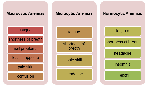
Clinical manifestations have to be properly identified because they may help patients recognize current health problems and ask for professional help. Despite its type, anemia’s main symptoms usually include fatigue, loss of energy, shortness of breath, and regular headaches, as shown in Figure 1 (Nagao & Hirokawa, 2017). Pale skin is also the reason to suspect anemia in a patient (Braunstein, 2019).
Iron deficiency may provoke hunger and a wrong nail construction, and vitamin B12 deficiency is characterized by a poor or lost sense of touch and dementia. RBC destruction results in abdominal pain, seizures, insomnia, chest pain, and the change of skin color. In children, attention is paid to the temperature of hands and feet because cold hands when the temperature of the body is normal or high is a sign of anemia and a call to action.

Conclusion
In total, the evaluation of anemia shows how complex this condition is for humans and their well-being. Although it is not a disease but a condition, its importance cannot be neglected because it contributes to the development of new health problems and complications. Macrocytic anemias due to vitamin B12 or folate deficiency are dangerous for people if no treatment is taken. Microcytic anemias because of iron deficiency can be inborn or acquired influence the growth of the body. Normocytic anemias are connected with inflammations and congenital problems. All these types may be properly diagnosed, treated, and prevented as soon as patient-doctor cooperation begins.
References
Adamu, A. L., Crampin, A., Kayuni, N., Amberbir, A., Koole, O., Phiri, A.,… Fine, P. (2017). Prevalence and risk factors for anemia severity and type in Malawian men and women: Urban and rural differences. Population Health Metrics, 15(1). Web.
Alder, L., & Tambe, A. (2019). Acute anemia. Web.
Braunstein, E. M. (2019). Evaluation of anemia. Web.
Chaparro, C. M., & Suchdev, P. S. (2019). Anemia epidemiology, pathophysiology, and etiology in low-and middle-income countries. Annals of the New York Academy of Sciences, 1450(1), 15-31.
Nagao, T., & Hirokawa, M. (2017). Diagnosis and treatment of macrocytic anemias in adults. Journal of General and Family Medicine, 18(5), 200-204.