Introduction
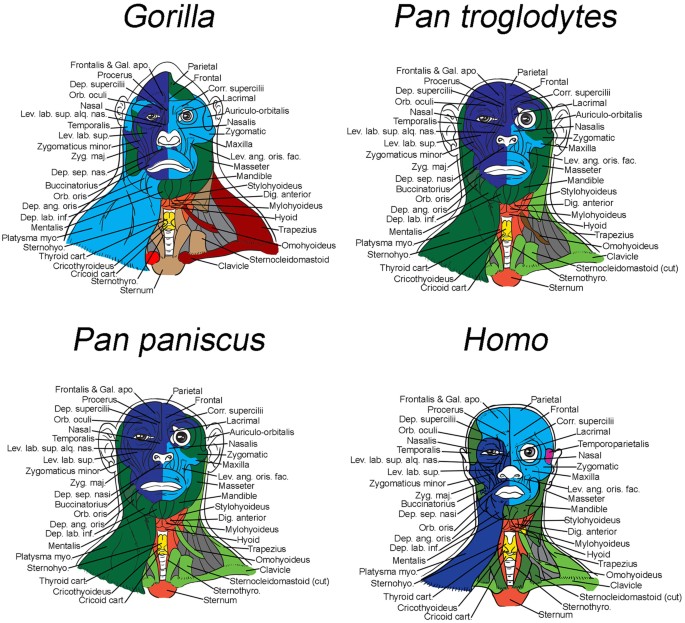
The muscles of the head and neck (Fig. 1) are divided into two groups: masticatory and mimic muscles. In some cases, they function together for articulating speech, chewing, swallowing, and yawning. The masticatory muscles are four paired muscles located on the sides of the skull. They all start on the bones of the skull and attach to the lower jaw, setting it in motion (Bordoni & Varacallo, 2022). The masticatory muscle begins from the lower edge of the zygomatic bone, the zygomatic arch; attaches to the masticatory tuberosity of the outer surface of the lower jaw. She raises the angle of the lower jaw.

Discussion
The temporal muscle begins from the temporal surface of the frontal bone, the parietal bone, the scales of the temporal bone (temporal fossa), the large wing of the sphenoid bone, the temporal fascia; attaches to the coronal process of the lower jaw. It raises the lower jaw (the biting muscle); the posterior tufts pull the jaw back. The medial pterygoid muscle begins from the pterygoid fossa of the pterygoid process of the sphenoid bone (Georgakopoulos & Lasrado, 2021). It attaches to the pterygoid tuberosity of the inner surface of the lower jaw. The medial pterygoid muscle raises the angle of the lower jaw.
The lateral pterygoid muscle begins from the suspensory crest of the large wing of the sphenoid bone, the outer surface of the lateral plate of the pterygoid process. It attaches to the neck of the lower jaw, the intra-articular disc and the capsule of the temporomandibular joint. With unilateral contraction, it shifts the jaw in the opposite direction, and with bilateral contraction, the lower jaw moves forward (Abuhaimed et al., 2022). Facial muscles are located under the skin, start from the bones of the skull and are woven into the skin. During contraction, the skin is shifted, changing its relief, forming facial expressions.
The neck muscles are topographically anatomically divided into superficial, medium, and deep. The superficial muscles are the subcutaneous muscle of the neck and the sternocleidomastoid muscle. The subcutaneous muscle of the neck is located directly under the skin, covering the entire front surface of the neck. It tightens the skin; pushing it forward, the muscle promotes the expansion of veins and the outflow of blood from the head (Powell et al., 2022). The subcutaneous muscle begins from the thoracic fascia, the skin of the upper chest at the level of the second rib (Bordoni & Varacallo, 2019). It attaches to the chewing fascia, the edge of the lower jaw, the corner of the mouth. The subcutaneous muscle of the neck pulls the corner of the mouth down, pulls the skin of the neck, prevents compression of subcutaneous veins.
Conclusion
The middle, or muscles of the hyoid bone, include: the muscles lying above the hyoid bone lie between the lower jaw and the hyoid bone. They are part of a complex apparatus, including the lower jaw, hyoid bone, larynx, windpipe, and play an important role in the act of articulate speech. Deep muscles include lateral muscles attached to the ribs. The anterior rectus muscle of the head starts from the anterior surface of the lateral mass of the atlas (Flynn & Vickerton, 2020). It attaches to the lower surface of the basilar part of the occipital bone. The lateral rectus muscle of the head begins from the transverse process of the atlas; it attaches to the lower surface of the jugular process of the occipital bone. It tilts his head to the side.
References
Abuhaimed, A. K., Alvarez, R., & Menezes, R. G. (2022). Anatomy, head and neck, styloid process. National Journal for Biotechnology Information, 40(13), 942–945.
Bordoni, B., & Varacallo, M. (2022). Anatomy, head and neck, sternocleidomastoid muscle. National Journal for Biotechnology Information, 42(2), 1–7.
Bordoni, B., & Varacallo, M. (2019). Anatomy, head and neck, temporomandibular joint. Europe PMC, 234(8), 540–547.
Flynn, W., & Vickerton, S. (2020). Anatomy, head and neck, larynx cartilage. Europe PMC, 382(21), 1941– 1960.
Georgakopoulos, B., & Lasrado, S. (2021). Anatomy, head and neck, inter-scalene triangle. National Journal for Biotechnology Information, 36(6), 397–402.
Powell, V., Esteve-Altava, B., Molnar, J., Villmoare, B., Pettit, A., & Diogo, R. (2018). Primate modularity and evolution: First anatomical network analysis of primate head and neck musculoskeletal system. Scientific Reports, 8(2341), 15–43.