Introduction
The skeletal component of the head that maintains the face and covers the brain is called the cranium or skull. It is further separated into the cranial vault and the facial bones. The facial bones hold the lower and upper jaw teeth, enclose the eyes, form the nasal cavity, and reinforce the facial tissues. The middle and inner ear components are housed inside the rounded brain case, which also encloses and safeguards the brain. The mature skull comprises 22 separate bones, 21 immovable and fused to form a single unit (Anderson et al., 2022). The lower jaw is the only bone in the skull that can be moved and is the 22nd bone.
Discussion
The facial bones that make up the anterior skull serve as the skeletal framework for the eye and other facial features. The apertures of the nasal cavity and orbit dominate this view of the cranium. The orbit forms the bony prominence in which the eyeball and the muscles that move it and raise the top eyelid are situated. The supraorbital margin refers to the outside edge of the anterior orbit. A little opening known as the supraorbital foramen can be found close to the supraorbital margin’s midpoint.
The nasal septum divides the nasal cavity, which is located inside the nasal region of the skull, into two halves. The vomer bone makes up the bottom section of the nasal septum, while the transverse plate of the ethmoid bone forms the top portion (Jamil & Callahan, 2022). The nasal canal takes a triangular shape, with a wide inferior area that narrows to a smaller superior space on either side. The inferior nasal concha, a separate skull bone, is the largest of the two bony plates on the lateral wall of the nasal cavity. The medial nasal concha, which comprises a portion of the ethmoid bone, is situated directly above the inferior concha (Lipsett & Khalid Alsayouri, 2022). The superior nasal concha is the ethmoid bone’s third bony plate component. Above the middle concha, it is considerably smaller and hidden from view.
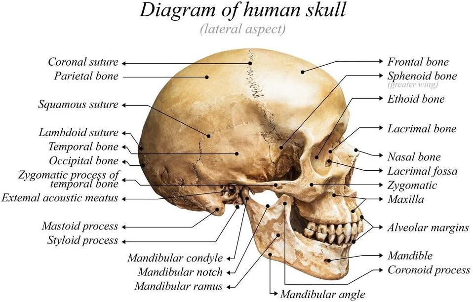
The massive, rounded brain case, located above, and the mandibles, located below, are the main features of a view of the lateral skull. The bone bridge known as the zygomatic arch divides these regions. The bony arch that runs from the cheek region upward from the ear canal is called the zygomatic arch, located on the skull’s edge. The zygomatic process of the temporal bone, which extends forward from the temporal bone, and the temporal process of the zygomatic bone, which is shorter and farther back, join to produce it. The zygomatic arch gives rise to one of the main muscles that lift the jaw upward while biting and eating. The temporal fossa is a small area located on the lateral aspect of the braincase, just above the zygomatic arch (Singh & Varacallo, 2022). Another area known as the infratemporal fossa is located deep in the vertical region of the jaw, underneath the zygomatic arch. The infratemporal and temporal fossa have muscles that work with the jaw to chew.
Conclusion
A suture is an immovable connection that connects two nearby skull bones. Thick, fibrous connective tissue fills the little space between the bones, holding them together. The lengthy sutures that connect the bones of the skull are not straight; rather, they wind in an asymmetrical, tightly twisted pattern. The sagittal and coronal sutures are the two lines of sutures that may be observed on the top of the skull (Russell & Russell, 2021). Across the cranium’s coronal plane, the coronal suture extends from side to side. The left, right, and right parietal bones are connected to the frontal bone through this joint. The sagittal suture runs parallel to and posterior to the coronal suture all along the midline at the anterior portion of the skull, where the left and right parietal bones are joined by it. The sagittal joins the lambdoid suture in the skull’s rear, where it ends. From its intersection with the sagittal suture, the lambdoid suture runs laterally to either side and downwards.

References
Amazon (2022). Laminated human skull diagram anatomy educational chart poster dry erase sign 18×12: Industrial & scientific. Amazon.com. Web.
Anderson, B. W., Kortz, M. W., & Kharazi, A. (2022). Anatomy, head and neck, skull. Nih.gov; StatPearls Publishing. Web.
Jamil, R. T., & Callahan, A. L. (2022). Anatomy, sphenoid bone. Nih.gov; StatPearls Publishing. Web.
Lipsett, B. J., & Khalid Alsayouri. (2022). Anatomy, head and neck, skull foramen. Nih.gov; StatPearls Publishing. Web.
Russell, W. P., & Russell, M. R. (2021). Anatomy, head and neck, coronal suture. Nih.gov; StatPearls Publishing. Web.
Singh, O., & Varacallo, M. (2022). Anatomy, head and neck, frontal bone. Nih.gov; StatPearls Publishing. Web.