Abstract
Anencephaly is a serious, inherent defect of the central nervous system that leads to the deformity of the brain and the associated tissues. Its main causes are linked to folate deficiency during pregnancy, genetic abnormalities and environmental factors. Statistics show that most anencephalic babies die prematurely, during birth or, in rare cases, a few months after birth. The diagnosis of the disorder entails performing ultrasonography and testing for biological markers such as maternal serum alpha-fetoprotein in the first and second trimesters. Elective abortions may be recommended for parents with anencephalic fetuses. No treatment options exist for the disorder, which causes immense emotional trauma to the concerned parents. Genetic counseling is recommended for parents with anencephalic babies to predict future chances of recurrence of the disorder and to help the parents prepare adequately through folic acid supplementation.
Description of Anencephaly
Anencephaly is a severe developmental flaw of the central nervous system that leads to the deformity of the brain and the cranial vault (Fleurke‐Rozema et al., 2015). The cerebrum and cerebellum may be nonexistent or diminished in size (Reddy & Ramanappa, 2016). However, the hind section of the brain is usually available. The terms meroanencephaly and holoanencephaly may be used to denote the magnitude of the cranial deformity. However, these terms do not foretell the gravity of the condition. Anencephaly is a disorder that falls into a category of disorders referred to as neural tube defect (NTD) spectrum (Reddy & Ramanappa, 2016). This flaw arises due to the failure of the neural tube to seal in the initial three to four weeks of development thereby causing fetal loss, stillbirth, or newborn mortality. In the usual development of a human embryo, the neural plate develops about 18 days following fertilization. The neural cover folds inwards along the length of the midplane of the embryo leading to the creation of the neural channel in the course of the fourth week of maturity. The sealing of the neural groove from the midline to both ends leads to the formation of the neural tube. This process terminates on the 24th day of pregnancy for the cranial end and the 26th day for the tail end. Any interference with the conventional closure of the neural groove produces a neural tube defect such as anencephaly, which happens when neural tube fails to close on the cranial end.
Being a neural tube defect, anencephaly keeps to a multifactorial model of transmission, which entails the association of several genes in addition to environmental factors. However, the precise genes and environmental factors involved have not been well elucidated. Certain instances of the disorder arise because of a chromosome aberration or multifarious developments embroiling imperfections of distinct genes. One gene implicated in the disorder is methylenetetrahydrofolate reductase (MTHFR), which is important in the metabolism of folic acid. Another gene known as VANGL1, which is a complex protein that is responsible for membrane-associated signaling, has also been shown to have a positive correlation with neural tube defects (Reddy & Ramanappa, 2016). Disturbances of the amniotic skin have also been implicated in anencephaly.
A number of environmental factors have been reported to facilitate the closing of the neural tube hence preventing the disorder. For example, folic acid and other related folates are known to prevent the disorder (Youngblood et al., 2013). However, substances such as folate antimetabolites, the presence of fungal toxins (mycotoxins) in corn products, and arsenic act as stressors that elevate the chances of NTDs such as anencephaly. Additionally, the health status of the mother plays a vital role. For example, disorders such as diabetes, obesity and hypothermia during pregnancy increase the risk of anencephaly in the developing fetus.
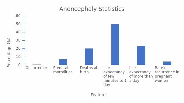
According to the Centers for Disease Control and Prevention, three out of 10,000 pregnancies have anencephaly in the U.S., which translates to an incidence rate of 0.001% (Centers for Disease Control and Prevention, 2016). Other studies report that about 7% of babies with anencephaly die in the course of pregnancy while 20% succumb during birth. 50% of these babies can live between a few minutes and one day while 23% can survive for more than one day (Jaquier, Klein, & Boltshauser, 2006). It has also been reported that more female babies than male babies are born with the condition.
Table on Anencephaly Statistics.
Signs and Symptoms of Anencephaly
Anencephaly is usually evident at birth due to the lack of the cranial vault and sections of the cerebrum and cerebellum. The face of the affected child may have all the facial characteristics and look normal. In rare cases, the cranial laceration is concealed by skin. However, it is left exposed in most cases. The concealing of the wound by skin complicates the process of prenatal diagnosis through the detection of a distinct protein known as maternal serum alpha-fetoprotein. Most babies with anencephaly are born dead (stillborn). Unprompted abortions in the course of pregnancy are also quite common. Mothers with anencephalic fetuses may have elevated levels of maternal serum alpha-fetoprotein (MSAFP) as well as excessive fluid in the amniotic sac.
How Anencephaly Affects the Body
Anencephaly is fatal in all instances due to the acute brain abnormality that characterizes the disorder. The brain plays a substantial role in regulating and directing most physiological processes. Consequently, a large number of anencephalic fetuses are aborted extemporaneously or stillborn. Anencephalic newborns have minimal chances of survival. Therefore, they tend to die within the early neonatal stage. It is presumed that children with anencephaly do not have the ability to see, hear or feel pain. However, families with anencephalic children report contrary opinions. These observations can be attributed to the differing extents of brain growth in various children. In some children, it may be possible to carry out normal physical processes expected of a child including feeding and crying. Some babies may also display the ability to respond to pressure and brightness.
Diagnosis and Identification of Anencephaly
Anencephaly can be diagnosed during the gestation period through ultrasonography. Ultrasonography, which is also known as medical ultrasound, is a diagnostic imaging method that employs high-frequency sounds to generate images of the inner organs and tissues (Fleurke‐Rozema et al., 2015). A gadget referred to as a transducer changes electrical current into sound waves that are directed into the tissues in the body. Sound waves recoil from the internal structures and are mirrored to the transducer that changes the waves into electrical signals. A computer transforms the tessellation of electrical signals into an image that is visualized on a screen. The image can also be captured on tape or film. It is advisable for a mother to avoid eating and drinking for a few hours prior to the test. This method is cost-friendly, non-invasive and is considered safe during pregnancy.
High levels of maternal serum alpha-fetoprotein indicate the possibility of anencephaly, which is confirmed by ultrasonography. MSAFP testing in the second trimester is a valuable tool for the diagnosis of most anencephaly instances in women with family histories of the disorder as well as those without family histories. The test is accurate between the 16th and 18th weeks of pregnancy and is suggested for diabetic women, those with a family record of birth flaws, women of advanced age, and those who have been exposed to harmful drugs during pregnancy. The levels of this protein change throughout pregnancy from 0.20 ng/mL to approximately 250 ng/mL by 32 weeks hence the need to date pregnancies precisely. Elevated levels of MSAFP are indicative of neural tube flaws and esophagus deformities. Conversely, low levels may indicate chromosomal aberration or Down’s syndrome.
Another useful test is the amniotic alpha-fetoprotein (AFAFP) screening. This test is performed anywhere from three to six months of gestation. An open neural tube in the fetus causes the protein to seep into the amniotic fluid thus leading to abnormally high levels of AFP. The AFP spreads to the maternal circulation and can be detected in the serum. In some cases, there may be false positive results. However, an additional test for acetylcholinesterase (ACHE) is performed to find an accurate diagnosis. ACHE tests are usually positive in anencephaly.
Laboratory tests cannot be performed after birth. Instead, cytogenetic tests are usually done to rule out trisomy 13 and lop-sided structural chromosome aberrations. However, ossification of the cranial vault is not reliably evident before the first twelve weeks of pregnancy are over. Therefore, ultrasonography should not be used in the diagnosis of anencephaly earlier than twelve weeks.
Treatments, Surgeries, and Therapies
The gravity of anencephaly does not encourage gallant stratagems to prolong the infant’s existence. Instead, the medical care team needs to concentrate on offering a compassionate atmosphere where the family can accept the diagnosis and prepare for their loss. Emotive support and grief counseling are needed for all affected families. Nevertheless, families that have had adequate time to take in the news of the diagnosis may appear prepared, but they must also be allowed to mourn. It may be necessary to include relatives, close acquaintances and religious leaders in the support system.
Following childbirth, families may wish to gain closure by carrying the affected baby and capturing a few moments with the baby through pictures. In such instances, it may be helpful to cover the baby’s head with a suitable piece of clothing. However, certain families may wish to observe the actual laceration. Medical providers should allow such parents to see the malformation to aid in dismissing conceptual images that could be exaggerations of the real image. Additionally, allowing the family to have intimate contact with their baby is useful in confirming the facts provided by the health experts thus helping the families in coming to terms with their loss.
Some couples may opt to proceed with the pregnancy even after the diagnosis of anencephaly. In such cases, it may be wise to discuss the possibilities of premature labor, polyhydramnios, late commencement of labor and other probable complications.
Alternative or Complementary Treatments
Pregnancy Care
All patients with prenatal diagnoses of anencephalic fetuses need to be referred to a care provider with adequate skills in conveying grave information. The provider should be knowledgeable about the chances of recurrence, preclusion, and screening for future pregnancies. An expert maternal fetal medicine physician can provide the required information in the absence of geneticists or genetic counselors. Details concerning the management of a current pregnancy should be discoursed in such consultations.
How a given pregnancy is handled following the diagnosis of anencephaly is influenced by how far the pregnancy has advanced before the diagnosis is made. Pregnancy termination is a possible option at pre-viable gestational ages (Johnson et al., 2012). However, the definition of the upper bound of the gestational age for pregnancy termination varies depending on the area of jurisdiction as well as the expertise of the medical personnel charged with conducting the procedure.
In instances where the parents disagree with pregnancy termination or when the pregnancy has already progressed to a sustainable gestational age, the treatment considerations change. It becomes necessary to decide whether or not to induce labor and the gestational age for labor induction. In such pregnancies, the usual bodily strains associated with gravidity are worsened by the psychological trauma of carrying a fetus with a deadly birth flaw or other medical conditions. Therefore, most doctors see it fit to induce labor.
Newborn resuscitation efforts, procedures and their costs should also be discussed before labor, planned and documented. The concerned delivery nurses, obstetricians and neonatal assistants should be notified of the patient’s wishes for her child. As already described, anencephaly is a severe disorder and the affected babies do not live beyond one month if born alive. Therefore, medical procedures to prevent premature delivery or cesarean delivery are not rational choices in such pregnancies.
Consultations
Once a couple has received an anencephalic diagnosis, it is vital for them to schedule an appointment with a geneticist. The sessions with the genetic expert will equip the parents with useful information regarding the disease, and will include how to ensure that the condition does not recur in the future as well as the available alternatives for testing and identifying the condition. In an ideal situation, the services of a genetic counselor should be sought prenatally and the following duration until the end of the pregnancy. Genetic counselors provide advice on how to get through the intricate psychosocial problems that families often have after receiving news that their babies have anencephaly.
Diet
It has been reported that the chances or the recurrence of anencephaly are 4% in mothers who have previously had babies with the condition. To prevent future cases, taking folic acid tablets as well as foods augmented with folate before trying to conceive and in the course of forthcoming gravidities is suggested (Youngblood et al., 2013). Adequate folates cannot be obtained from diet alone. Therefore, a daily intake of 400 µg folic acid is recommended (Youngblood et al., 2013).
Medications
Regrettably, no treatment is available for a child with anencephaly. However, the baby and his family can still receive moral support. Perinatal hospice care, which is usually offered in many hospitals provides support for these families from pregnancy, delivery, and death by honoring the memories of the baby. Perinatal hospice is a frame of mind that specializes in providing care to the baby and his parents in a loving and honorable manner.
Conclusion
Anencephaly is a congenital defect with fatal effects on the fetus due to the abnormality of brain development. Even though no treatment exists, families with affected babies can receive adequate counseling to help them cope with the loss of their babies.
References
Centers for Disease Control and Prevention. (2016). Facts about anencephaly. Web.
Fleurke‐Rozema, J. H., Leijden, L., Kamp, K., Pajkrt, E., Bilardo, C. M., & Snijders, R. J. M. (2015). Timing of detection of anencephaly in The Netherlands. Prenatal Diagnosis, 35(5), 483-485.
Jaquier, M., Klein, A., & Boltshauser, E. (2006). Spontaneous pregnancy outcome after prenatal diagnosis of anencephaly. BJOG: An International Journal of Obstetrics and Gynecology, 113(8), 951-953.
Johnson, C. Y., Honein, M. A., Dana Flanders, W., Howards, P. P., Oakley, G. P., & Rasmussen, S. A. (2012). Pregnancy termination following prenatal diagnosis of anencephaly or spina bifida: A systematic review of the literature. Birth Defects Research Part A: Clinical and Molecular Teratology, 94(11), 857-863.
Reddy, J., & Ramanappa, M. V. (2016). Anencephaly and its associated anomalies in antenatal scans, Journal of Evidence Based Medicine and Healthcare, 3(23), 1033-1035.
Youngblood, M. E., Williamson, R., Bell, K. N., Johnson, Q., Kancherla, V., & Oakley, G. P. (2013). 2012 Update on global prevention of folic acid–preventable spina bifida and anencephaly. Birth Defects Research Part A: Clinical and Molecular Teratology, 97(10), 658-663.