Abstract
The Down’s syndrome is the problem that may occur in a number of reasons. The main purpose of the research is to identify the authors who considered the problem of Down’s syndrome causes and analyze the results of the research conducted by those scholars. The main result of the current research is that it was enumerated a number of reasons which may be the causes for the Down’s syndrome appearance. The Down’s syndrome is mostly caused by an error during cell division process that is resulted in an extra genetic material. This extra genetic material creates 47 chromosomes in human organism instead of 46, a norm. The most frequently occurred consequences of those changes are lack of cognitive, physical and speech development. It was also possible to identify the types of Down’s syndrome, namely, mosaic, trisomy 21, and translocation.
Introduction
Down’s syndrome is a disorder which is caused by the addition of an extra genetic material that is associated to causing delays in the development of a child in this condition. Down’s syndrome was named after a British physician, John Langdon Down; he is the one who described the symptoms first in 1886. He published an essay illustrating different young children who suffered from mental retardation. While working in an asylum in England, Down found a distinction between children who are cretins and those he called Mongolods. Mongoloidism, a term he used because these children had similar characteristics as those of people from Mongolia who had arrested development. An extensive amount of research was conducted after Down’s findings. Though out this paper, different studies will be discussed to help us understand what causes a person to be born with Down’s syndrome; the paper will also analyze the relationship between maternal serum level of human placental growth hormone (HPGH) and fetal Down syndrome.
General Information on Down’s syndrome
Down’s syndrome has an occurrence rate of one out of 600 live births (5,500 lives annually) in nearly all races and across the economic classes . The occupational categories, still, influence people’s ability to give birth to a live-child with Down’s syndrom . It is proved that the chances to give birth to a baby with Down’s syndrome increase with the increase of mother’s age. Thus, 35-year-old woman has 1:385 chances to give birth to a baby with Down’s syndrome, while the risk of 40-year-old woman is 1:30 . There are a great number of different tests which are able to identify the Down’s syndrome on different terms of pregnancy.
It was considered with the help of Serum, Urine and Ultrasound Screening Study (SURUSS), the First and Second Trimester Evaluation of Risks (FASTER) Trial and the Serum Biochemistry and Fetal Nuchal Translucency Screening (BUN) that the first trimester serum screening is the safest and allows identify the problem on the earliest terms . Three forms of Down’s syndrome are known to exist and they include translocation, trisomy 21, and mosaic. In trisomy 21, every cell in the body has the extra chromosome 21. Tristomy 21 has 97% of the population suffering from the disease. Translocation type of Down’s syndrome is the much more rarely met, it is a chromosomal abnormality that comes across in 4% of all cases of the disease. The mosaicism is the rarest and covers only 1% of the cases .
The problem of non-disjunction
It is impossible to dwell upon Down’s syndrome and its reasons without turning to the problem of non-disjunction. The main literally meaning of non-disjunction is “not coming apart”. It is the process when chromosome pairs fail to separate properly during cell division. To understand the Down’s syndrome occurrence is impossible without the understanding of the reasons for non-disjunction. There is a hypothesis that the main reason for non-disjunction is a cascading event, which is the subsequence of “hormonal imbalance, sub-optimal micro-vasculature around the ovarian follicle, reduced blood flow, increased carbon dioxide and lactic acid inside the follicle, decreased pH in the oocyte, reduced mitotic spindle size, spindle displacement and non-disjunction” . A number of the research was conducted on the problem of reasons for non-disjunction that causes the Down‘s syndrome. The following conclusions were obtained, the non-disjunction may be the result of secondary non-disjunction (when one of the parents was trisomy 21), translocation, the presence of the specific gene of non-disjunction in human organism, or the environmental effect that should not be omitted .
Types of Down’s syndrome
Translocation

Having considered some reasons for non-disjunction that may result in Down’s syndrome, it is possible to shift to the problem of three types of the disease, translocation, trisomy 21, and mosaic, in detail. Turning to translocation (Pic.1), it is necessary to mention that those who suffer from problems caused by translocation have “2-way exchange of material between 2 nonhomologous chromosomes, with no net gain or loss of genetic material” . Translocation is not actually a change of the chromosomal material, but it is the change of the genetic material location. The commonly occurred case of the translocation is the exchange of the genetic material between “arms of two heterologous chromosomes” . Being one of the most commonly met balanced structural chromosomal abnormality, translocation occurs in 1 one per 1175 cases. To the point, the unbalanced structural chromosomal abnormality in humans occurs in about 0.04% cases but it is an extremely significant factor in morbidity and mortality cases .
Trisomy 21
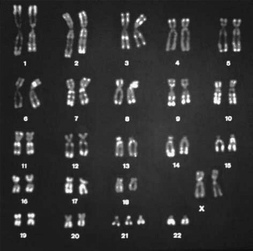
As it was mentioned above, trisomy 21 (Pic. 2) is one of the reasons for the Down’s syndrome occurrence. Being one of the most frequently befallen, trisomy 21 takes place in 1 of 700 cases without the relation to any specific ethnic group. Trisomy 21 is caused by the meiotic errors that occur in egg and involve an extra maternal chromosome in 90% of cases of Down’s syndrome presence. Still, trisomy 21 is the cause of a number of other problems which attend the Down’s syndrome, such as “congenital heart disease, duodenal.
A person suffers from Down stenosis or atresia, imperforate anus, syndrome. Hirschprung disease, muscle hypotonia, immune system deficiencies, increased risk of childhood leukemia and early onset Alzheimer’s disease” . Some research was conducted on the main effects of trisomy 21. It is an undeniable truth that gene changes in human organism are extremely harmful and negatively affect people’s mental and physical health. Thus, there are some facts that may not be omitted. The genetic changes that occur in human organism due to trisomy 21 affect the cancer development and prevent it. The research was conducted for about 50 years proved that the gene dosage of the trisomy 21 protectively affects cancer cells and especially Ets2 gene dosage that is responsible for the tumorigenesis .
Mosaic

Mosaicism is one of the rarest types of Down’s syndrome. As it was stated above it covers 1% of disease cases in the world. Mosaicism is characterized by the possession of trisomy 21 but with specific characteristics when there may occur both normal and trisomic cell lines. Some sources report that “mosaicism is caused by the loss of the extra chromosome of a trisomic zygote in 80% of all cases and that the remaining 20% probably result from mitotic non-disjunction of an euploid zygote” .
The main peculiarity of the mosaic Down’s syndrome is that people possessing it on the genetic level may either have the intellectual disabilities of omit them. It was proved via the research that people with mosaic Down’s syndrome may have strong signs on the intellectual level, the other patients may get just some of the signs and the last group may omit any characteristics of the disease and show absolutely normal IQ level . The more detailed results on the comparative analysis of children with trisomy 21 and mosaic Down’s syndrome may be presented. Thus, children with trisomy 21 managed to get from 18 to 75 range of IQ level with the mean of 52, while those with mosaic modification of the disease were able to receive from 43 to 92 and the mean IQ is 67 .
There are various signs and symptoms that are characteristic of a person suffering from Down syndrome. The asynchrony in the development of those with Down’s syndrome is notable. It is significant to remember that children with Down’s syndrome have communicational difficulties. They prefer to use gestures and signs while communication to speaking {/867b}. Furthermore, due to weak and slow development, children are unable to lead the normal communication with others. The problem is also supported with the differences in physical development. Those suffering from Down’s syndrome start walking later. It is the result of the delays in motor development, “as well as the development of abnormal compensatory movement patterns related to abnormal foot, knee, hip and ankle kinetics” .
The difficulties in communication and differences in physical abilities create a number of complications while the collaboration between children with Down’s syndrome and their unaffected siblings. The prejudiced attitude of the society to people with Down’s symptom leads to their labeling as stigmatized identity. The situation is also worsen by the fact that those who suffer form the disease consider themselves retarded and different, their self-esteem is rather low and results in negative personal evaluation. The negative attitude of people with Down’s syndrome to their personality leads to isolation, and, as a result, to the social unawareness .
The society, knowing not much about people with Down’s syndrome, tries to avoid them and this leads to some hostility. It is also notable to mention another effect, when people with Downs’ syndrome aggressively react to the society. People who suffer from the disease were interviewed and followed and the reaction on some situations was considered. IT may be concluded that the research showed that the aggressive reaction of people with Down’s syndrome was three times lower that that of people with learning disabilities, still it was. It is also crucial to mention that neither epilepsy no autism were the reasons for this aggression . The research of disease suffering people’s aggression should be continued as the mentioned study did not give specific answers; it just excluded two reasons for aggression of people with Down’s syndrome and put the problem higher than it was. In the relation to the behavior of people with Down’s syndrome, the research shows that the people with the disease are flexible in their behavior .
The relationship between maternal serum level of human placental growth hormone and fetal Down’s syndrome
It must be noted that the growth hormone (GH) is produces by human placenta. Before turning to the detailed research in the question, it is significant to understand the role of the human placental growth hormone (hPGH) in the extravillous cytotrophoblast (EVCT) invasiveness. This role is significant as according to the cellular model, hPGH is effective and influence the increase of EVCT invasive potential . Returning to the question of the significance of the placental GH on the fetal growth, it is important to know that different kinds of research prove the influence of placental GH on the fetal growth. Such conclusion was possible with the understanding that placental GH is interrelated with fetal growth. At the same time, there were no direct signs of IGF-I influence in fetal growth. Some logical conclusions may create a hypothesis that IGF-I influenced fetal growth indirectly, via placental size and function affection .
The suggestion was made that maternal serum level of hPGH may play a significant role in identifying the problems that deal with the chromosomal anomalies while pregnancies. The maternal serum level of hPGH may be a kind of marker that could help identify the signs of Down’s syndrome while screening . Frendo J.L, Vidaud M, Guibourdenche J, et al. made an attempt to explain why the maternal serum level of hPGH is a potential marker for chromosomal anomalies identification. They are sure that the Down’s syndrome is identified by means of a number of signs which are connected directly to the placenta development that is resulted in the hormones’ influence.
First, the syncytiotrophoblast is the core layer in the placenta that is responsible for a number of processes while pregnancy. This syncytiotrophoblast layer is the surface one and is seen best of all. The significance of this layer is that it is formatted with abnormities in case of Down’s syndrome occurrence. Furthermore, the decrease “in the production of pregnancy-specific polypeptide hormones hPGH as well] by the placenta in Down’s syndrome” is observed . All the facts mentioned above prove that the relation between maternal serum level of human placental growth hormone and fetal Down’s syndrome is on the high level.
Maternal serum screening for Down’s syndrome
There are several types of screening which may be helpful while identifying the Down’s syndrome sighs while pregnancy. Fetal nuchal translucency method is one of the most effective one in the chromosomal abnormalities identification while screening. The increased thickness in fetal nuchal translucency is the sign for the Down’s syndrome occurrence. Still, the gestational age should be taken into account as its ignoring may result in false positive rates. Furthermore, the use of the mixture of methods may be a good opportunity to know for sure either the presence or the absence of the Down’s syndrome is observed in pregnancy .
The Down’s syndrome identification on the early terms of the pregnancy is important. There is an opinion that the early identification of the Down’s syndrome is unreliable and that the second trimester should be waited. Thus, this opinion is false. The research conducted among 4412 women showed the following (Pic. 3)
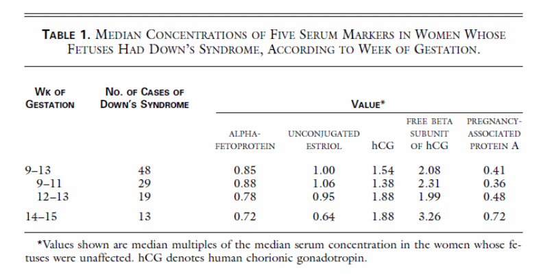
The table allows consider the concentration of different markers in women organism while pregnancy on the first and second trimesters. Some of the rates are higher, the others are lower . The level of serum concentration in women organism allows us to conclude that there is no need to wait till the second trimester and the identification of the Down’s syndrome may be provided on the first one. Palomaki et al. is still sure that the conclusion about either the presence or the absence of the Down’s syndrome should be provided on the second trimester by means of combining the of the first and second trimesters as the use of the markers combination may help state precise results .
Still, Mennuti and Driscoll still believe that the Down’s syndrome screening should be provided in the second-trimester on the general basis, while this does not exclude women’s desire to be screened on the first trimester of pregnancy . There is an idea to use the combination of the Downs’ syndrome, identification, the maternal serum test in the combination with ultrasonography . The combination of sonographic examination and maternal serum testing maximizes the benefits of Down’s syndrome identification, if it is present, no matter which succession is chosen .
It is significant to underline that women who were received positive Down’s syndrome results while the first screening are less likely to come thorough the other screening while the second pregnancy. Even the fact that the risk of Down’s syndrome possession is higher in those who were screen-positive for the first time, do not influence their decision to avoid screening while the second pregnancy . When the women have the negative rhesus blood group, the tests should be conducted twice and the combinations and analysis of the achieved results may be the final conclusion of whether the foetus is affected with Down’s syndrome or not .
The importance of screening on both, the first and the second trimesters is significant. Some medics stress that the screening on the first trimester helps not just in trisomy 21 identification, but also in checking the number fetal malformations . Driscoll and Gross ruin the prejudice that maternal age may be a good screening criterion stressing that the vast majority of children with Down’s syndrome appears in women who no older than 35 . Furthermore, the very system of the Down’s syndrome screening was invented in late 1980s – early 1990s. The change in the software capacities and other life qualities should be taken into account and the change of the monitoring parameters should be provided to reduce the screen-positive errors rates .
Crang-Svalenius, Dykes, and Jörgensen are sure that the fetal neuchal translucency should be conducted on the first trimester while the serum screening is more promising on the second trimester term . The screening in the middle of the pregnancy term should be conducted with the help of different maternal serum biochemical markers “maternal serum α-fetoprotein (MSAFP), human chorionic gonadotropin (hCG), unconjugated estriol, and, recently, dimeric inhibin A” .
Some scholars stress that the reason for the false positive screen for Down syndrome may be the high level of serum concentrations of hCG . Most medics identify the error rate of 5% for Down’s syndrome positive screen . It is significant that some women consider the role of the serum screening in a wrong way. Thus, some participants of the experiment consider it as the helping strategy “to protect the fetus instead of finding cases of untreatable birth defects” .
Discussion
The researches above have clearly indicated a common trend in showing the relationship between maternal serum levels of hPGH and foetal Down’s syndrome. The levels of hPGH at Down’s syndrome pregnancies have been shown to change in the second trimester of the gestation period . The three main types of the Down’s syndrome were identified, trisomy 21, translocation and mosaic. Though, the type of the Down’s syndrome does not influence the choice of the type of identification of the problem. It is also understood that the first trimester of pregnancy is the best time to start the Down’s syndrome risk assessment with the help of the combination of fetal neuchal translucency thickness and maternal age . Fetal neuchal translucency is considered to be one of the most reliable tests on the Down’s syndrome possession .
It is crucial to know that Down’s syndrome problem is one of the main prototypes for research of people’s aneuploidy . The translocation and mosaic syndromes are the types of trisomy which either replaces the chromosomes or changes their look. The change of the location of the trisomy 21 significantly influences the disease, but it still remains Down’s syndrome . The relationship between maternal serum level of hPGH and fetal Down’s syndrome is significant as the hPGH influences the placenta which, in its turn, creates some abnormalities which are easily shown on the screening and in case of Down’s syndrome may be easily considered.
Conclusion
There are some health conditions which have no cure and only require specialized care in order to reduce the patient’s suffering. They have been stigmatized in our societies therefore leaving the patients being locked at homes or caring centers. Thus, hospitalization may occur because of health problem reasons . Down’s syndrome which is also referred to as trisomy 21 is one of these conditions affecting our societies. Down’s syndrome occurs in 5,500 cases each year in nearly all races and across the economic classes . The condition is known to occur when there is an error during cell division process, this error is referred to as disjunction.
Normally, it occurs as a result of a fault in cell division called non-disjunction . As a result an extra genetic material is added either to the ovum or the sperm. During conception, the embryo acquires extra genetic material to have 47 chromosomes instead of the normal 46. It is this extra genetic material that causes lack of cognitive development, physical development and speech development. There are three common types of Down’s syndrome. These include mosaic, trisomy 21, and translocation. Much research has been carried out to find the relationship between maternal serum levels of hPGH and Down’s syndrome. The results are unanimous in that maternal serum levels of hPGH are increased in people with DS pregnancies. More research should be conducted in the future to provide the much needed knowledge of finding the cure for this syndrome. When this is done it will be a relief to parents and the children suffering from Down’s syndrome.
Bibliography
- Improved national prevalence estimates for 18 selected major birth defects–United States, 1999-2001. MMWR. Morbidity And Mortality Weekly Report. 2006; 54(51): 1301-1305.
- Khoshnood B, De Vigan C, Vodovar V, Bréart G, Goffinet F, Blondel B. Advances in Medical Technology and Creation of Disparities: The Case of Down Syndrome. American Journal of Public Health. 2006; 96(12): 2139-2144.
- Pinto S, Schub T. Down Syndrome. [serial online]. 2010; Available from: CINAHL Plus with Full Text, Ipswich, MA.
- Weisz B, Rodeck CH. An update on antenatal screening for Down’s syndrome and specific implications for assisted reproduction pregnancies. Human Reproduction Update. 2006; 12: 513-518.
- Pangalos C et al. Understanding the Mechanisms of Mosaic Trisomy 21, By Using DNA Polymorphism Analysis. American Journal of Human Genetics. 1994; 54(9): 473-481.
- Down syndrome. In: Strickland BB, ed. The Gale Encyclopedia of Psychology. Detroit: Gale; 2001: 193-194.
- Cuckle HS. Primary prevention of Down’s syndrome. International Journal of Medical Sciences. 2005; 2(3): 93-99.
- Read S. The distribution of Down’s syndrome. Journal of Mental Deficiency Research. 1982; 26 (Pt 4): 215-227.
- Dundar M, Caglayan A, Saatci C, Arslan K, Ozkul Y. Down syndrome like appearance with a novel de novo translocation t(6;21)(q21;q13). Indian Journal of Medical Research. November 2008; 128(5):666-668.
- Sommer C, Henrique-Silva F. Trisomy 21 and Down syndrome: a short review. Brazilian Journal Of Biology = Revista Brasleira De Biologia. May 2008; 68(2): 447-452.
- Sussan T, Yang A, Li F, Ostrowski M, Reeves R. Trisomy represses ApcMin-mediated tumours in mouse models of Down’s syndrome. Nature. 2008; 451(7174): 73-75.
- de A. Moreira L, San Juan A, Pereira P, de Souza C. A case of mosaic trisomy 21 with Down’s syndrome signs and normal intellectual development. Journal of Intellectual Disability Research. 2000; 44(1): 91.
- Fishier K, Koch R, Donnell G. Comparison of Mental Development in Individuals With Mosaic and Trisomy 21 Down’s Syndrome. Pediatrics. 1976;58(5):744.
- Toth A. Bridge of Signs: Can Sign Language Empower Non-Deaf Children to Triumph over Their Communication Disabilities?. American Annals of the Deaf. 2009; 154(2): 85-95.
- Whtt-Glover M, O’Neill K, Stettler N. Physical activity patterns in children with and without Down syndrome. Pediatric Rehabilitation. April 2006; 9(2): 1.
- Cunningham C, Glenn S. Self-awareness in Young Adults with Down Syndrome: I. Awareness of Down syndrome and disability. International Journal of Disability, Development & Education. 2004; 51(4): 335-361.
- Tyrer F, McGrother C, Thorp C, et al. Physical aggression towards others in adults with learning disabilities: prevalence and associated factors. Journal of Intellectual Disability Research. 2006; 50(4): 295-304.
- Didden R, Sigafoos J, Green V, et al. Behavioural flexibility in individuals with Angelman syndrome, Down syndrome, non-specific intellectual disability and Autism spectrum disorder. Journal of Intellectual Disability Research. 2008;52(6):503-509.
- Lacroix MC, Guibourdenche J, Fournier T, et al. Stimulation of Human Trophoblast Invasion by Placental Growth Hormone. Endocrinology. 2005; 146: 2434–2444.
- Chellakooty M, Vangsgaard K, Larsen T et al. A longitudinal Study of Intrauterine Growth and the Placental Growth Hormone (GH)-Insulin-Like Growth Factor I Axis in Maternal Circulation: Association Between Placental GH and Fetal Growth. Journal Clinical Endocrinology Metabolism. 2004; 89(6): 384-391.
- Baviera G, Carbone C, Corrado F, Mastrantonio P. Placental growth hormone in Down’s syndrome screening. Journal of Maternal-Fetal & Neonatal Medicine. 2004; 16(4): 241-243.
- Frendo JL, Vidaud M, Guibourdenche J, et al. Defect of Villous Cytotrophoblast Differentiation into Syncytiotrophoblast in Down’sSyndrome. Journal Clinical Endocrinology Metabolism. 2000; 85: 3700–3707.
- Wright D, Kagan KO, Molina FS, Gazzoni A, Nicolaides KH. A Mixture Model of Nuchal Translucency Thickness in Screening for Chromosomal Defects. Ultrasound Obstetrics Gynecology. 2008; 31: 376–383.
- Haddow J, Palomaki GE, Knight GJ. Josephine Williams, Wayne A. Miller, M.D., and Anthony Johnson, D.O. Screening of Maternal Serum for Fetal Down’s Syndrome in the First Trimester.
- Hellenic Endocrine Society. 1998; 338 (14): 955-962.
- Palomaki GE, Neveux LM, Knight GJ, Haddow JE, Pandian R. Maternal Serum Invasive Trophoblast Antigen (Hyperglycosylated hCG) as a Screening Marker for Down Syndrome during the Second Trimester. Clin. Chem. 2004; 50: 1804-1808.
- Mennuti MT, Driscoll DA. Screening for Down’s Syndrome — Too Many Choices?. NEJM. 2003; 349: 1471-1473.
- Wald N, Jenkins TM, Wapner RJ, Reynolds T, Zimmermann R, Wright E, Malone FD, D’Alton ME, Berkowitz RL, Watt H, Hackshaw A, Copel JA, Bahado-Singh RO. Integrated Screening for Down’s Syndrome. NEJM.1999; 341(7): 461-467.
- Pinette MG, Egan JFX, Wax JR, Blackstone J, Cartin A, Benn PA. Combined Sonographic and Biochemical Markers for Down Syndrome Screening. J Ultrasound Med. 2003; 22: 1185-1190.
- Rausch D, Lambert-Messerlian G, Canick, J Participation in maternal serum screening for Down syndrome, neural tube defects, and trisomy 18 following screen-positive results in a previous pregnancy. West J Med. 2000; 173(3): 180–183.
- Muhcu M, Mungen E, Dundar O, et al. Reliability of second trimester triple screening for Down syndrome in rhesus-negative women. Journal of Perinatology. 2007; 27(5): 268-271.
- Kagan K, Wright D, Baker A, Sahota D, Nicolaides KH. Screening For Trisomy 21 by Maternal Age, Fetal Nuchal Translucency Thickness, Free Beta-Human Chorionic Gonadotropin, and Pregnancy Associated Plasma Protein-A. Ultrasound. Obstetric Gynaecology. 2008; 31: 618–624.
- Driscoll DA, Gross S. Prenatal Screening for Aneuploidy. NEJM. 2009; 360: 2556-2562.
- Reynolds T M, Aldis J. Median parameters for Down’s syndrome screening should be calculated using a moving time-window method. Ann Clin Biochem. 2008; 45: 567-570.
- Crang-Svalenius E, Dykes A, Jörgensen C. Maternal serum screening for Down syndrome–opinions on acceptance from Swedish women. Scandinavian Journal Of Caring Sciences. 2003; 17(1):30-34.
- Lehavi O, Aizenstein O, Evans M, Yaron Y. 2nd-trimester maternal serum human chorionic gonadotropin and alpha-fetoprotein levels in male and female fetuses with Down syndrome. Fetal Diagnosis And Therapy. 2005;20(3):235-238.
- Abdul-Hamid S, Fox R, Martin I. Maternal serum screening for trisomy 21 in women with a false positive result in last pregnancy. Journal Of Obstetrics And Gynaecology: The Journal of the Institute of Obstetrics And Gynaecology. 2004; 24(4):374-376.
- Soergel P, Pruggmayer M, Schwerdtfeger R, Muhlhaus K, Scharf A. Screening for trisomy 21 with maternal age, fetal nuchal translucency and maternal serum biochemistry at 11-14 weeks: a regional experience from Germany. Fetal Diagnosis And Therapy. 2006; 21(3):264-268.
- Chiang H, Chao Y, Yuh Y. The maternal self in pregnant women undergoing maternal serum screening. Journal Of Clinical Nursing. 2007;16(6):1180-1185.
- Papadopoulou E, Sifakis S, Giahnakis E, et al. Human Placental Growth Hormone Is Increased in Maternal Serum in Pregnancies Affected by Down Syndrome. Fetal Diagnosis & Therapy. 2008;23(3):211-216.
- Baviera G, Carbone C, Corrado F, Mastrantonio P. Placental growth hormone in Down’s syndrome screening. Journal of Maternal-Fetal & Neonatal Medicine. 2004; 16(4): 241-243.
- Snijders RJ, Noble P, Sebire N, Souka A, Nicolaides KH. UK Multicentre Project on Assessment of Risk of Trisomy 21 by Maternal Age and Fetal Nuchal-Translucency Thickness at 10-14 Weeks of Gestation. Fetal Medicine Foundation First Trimester Screening Group. Lancet. 1998; 352: 343–346.
- Chang, TC. Antenatal screening for Down syndrome in New Zealand: Time for a national screening policy?. Australian & New Zealand Journal of Obstetrics & Gynaecology. 2006; 46(2):92-96.
- Korenberg J, et al. Down Syndrome Phenotypes: The Consequences of Chromosomal Imbalance. Proctor National Academy Science. USA. 1994; 91: 4997-5001.
- Shapiro, B. Whither Down Syndrome Critical Regions? Human Genetics. 1997; 99: 421-423.
- So S, Urbano R, Hodapp R. Hospitalizations of infants and young children with Down syndrome: evidence from inpatient person-records from a statewide administrative database. Journal of Intellectual Disability Research. 2008; 52(6): 503-509.
- Improved national prevalence estimates for 18 selected major birth defects-United States, 1999-2001. MMWR. Morbidity And Mortality Weekly Report. 2006; 54(51): 1301-1305.
- Ghosh D, Sinha S, Chatterjee A, Nandagopal K. A study of GluK1 kainate receptor polymorphisms in Down syndrome reveals allelic non-disjunction at 1173(C/T). Disease Markers. 2009; 27(2): 45-54.