Executive summary
Noninvasive cardiology refers to the diagnosis and therapy of a heart condition by external examination of the heart using techniques that enable the visualization of the heart tissues. Noninvasive cardiology was developed by researchers who were experimenting with ways to guide the catheters during heart examination. The researchers were mainly radiologists and they developed x-ray to take images of the heart indicating the location of the catheter. On further research, it was discovered and that the images revealed the anatomy of the heart and therefore information on the status of the heart could be gained by examining the images. This gave birth to noninvasive cardiology, and since then many more methods (ECG, PET, and CT scan) have been developed, and it is now possible to give a precise description of the heart condition using these techniques.
Noninvasive cardiology has impacted medicine in many different ways. The positive impacts include: Early detection of cardiovascular and heart diseases, provides a safer method for examination and treatment of heart ailments, is cost-effective, has a high specificity and reduces the cardiologist’s workload. However, the technology has its shortcomings too, the initial cost of installation of a noninvasive cardiology unit is very high. There is a lack of enough specialists in the field to run cardiology departments in hospitals. Lastly, operators and patients are at risk of cancer due to ionizing radiation and radioactive elements used in therapy and diagnosis.
Introduction
Invasive cardiology refers to the application of invasive techniques to treat heart diseases. These procedures entail the insertion of an instrument into the venous system to treat a condition. Noninvasive cardiology refers to the diagnosis and therapy of a heart condition by external examination of the heart using techniques that enable the visualization of the tissues of the heart. There are many forms of cardiovascular diseases, Cardiac failure (acute cardiac failure, congestive cardiac failure and chronic cardiac failure). This disease occurs due to insufficient cardiac output. Other diseases of the cardiovascular system include stenosis (narrowing of valve opening), hypertension, aneurysm, atheromas and myocardial infarctions.
Heart diseases have been found to be the major causes of death globally, according to the WHO:
- Heart attacks and strokes are major–but preventable–killers worldwide. Over 80% of cardiovascular disease deaths take place in low-and middle-income countries and occur almost equally in men and women. The cardiovascular risk of women is particularly high after menopause.
- Tobacco use, an unhealthy diet, and physical inactivity increase the risk of heart attacks and strokes.
- Cessation of tobacco use reduces the chance of a heart attack or stroke.
- Engaging in physical activity for at least 30 minutes every day of the week will help to prevent heart attacks and strokes.
- Eating at least five servings of fruit and vegetables a day, and limiting your salt intake to less than one teaspoon a day, also helps to prevent heart attacks and strokes.
- High blood pressure has no symptoms but can cause a sudden stroke or heart attack. Have your blood pressure checked regularly.
- Diabetes increases the risk of heart attacks and stroke. If you are a diabetic control your blood pressure and blood sugar to minimize your risk.
- Being overweight increases the risk of heart attacks and strokes. To maintain ideal body weight, take regular physical activity and eat a healthy diet.
- Heart attacks and strokes can strike suddenly and can be fatal if assistance is not sought immediately. (World health organization 2009)
Background
“The development of cardiology began as early as 18th century with the invasive techniques.”(Richards, 2010) The noninvasive cardiology technology was a culmination of many experiments by different groups working separately; many of them were radiologists who were looking for ways to improve invasive cardiology. The earliest research on cardiology can be traced back to 1711 when the first cardiac catheterization was by Stephen Hales. He positioned cardiac catheters into both ventricles of a living horse. “The first incidence of catheterization in humans was done by Werner Forssmann on himself in 1929. He made an incision into his left antecubital veins and inserted a catheter into his venous system. Using fluoroscopy, he guided the catheter into the right atrium.” ((Littmann, 2010) In the 1940s more methodical measurements of the hemodynamics of the heart were performed using catheterization. A major step in cardiac catheterization was made in 1958 by Charles Dotter who developed a technique (diagnostic coronary angiogram) that enabled the visualization of the coronary anatomy. This involved the injection of a tiny amount of contrasting radiographic agents which facilitated the production of radiographic films showing the coronary anatomy. In the 1950s the “cut down” procedure that involved the dissection of soft tissues to locate a vein or artery was phased out to pave way for a percutaneous approach. (Littmann, 2010) This technique is commonly referred to as the Seldinger technique and involves the puncturing of the vessel with a sharp hollow needle. A blunt sheath is then introduced into the vessel through the hollow needle. Catheters can then be passed through the sheath. Most of these early developments focused on invasive methods due to a lack of adequate technology. However, invasive cardiology had many contraindications; instances of hemorrhages were high and sometimes infections were passed to the patient. The challenges encountered led to the development of techniques which were to guide the catheters while inside the patient’s body. These techniques were meant to facilitate the visualization of the catheters in the patient’s body. However, it turned out that the techniques could also enable the visualization of the heart and hence direct examination.
The discovery and development of technologies like x-ray, electrocardiography (ECG) or cardiac ultrasound, computed tomography and radiotherapy have led to the successful development of non-invasive cardiology. Much progress has been witnessed since the first application of noninvasive techniques. This paper will seek to determine the impact of non-invasive cardiology Technology in medicine.
How Noninvasive Cardiology Technology (Nict) Works
Components
Noninvasive cardiology technology works via different systems whose aim is to diagnose or treat heart conditions. The most commonly used techniques include chest x-ray, Electrocardiogram (ECG), measurement of cardiac output using Positron emission tomographic (PET) myocardial perfusion and computed tomography. The techniques may be used in combinations to increase the reliability of the test results.
Techniques Involved in the Noninvasive Cardiology Technology
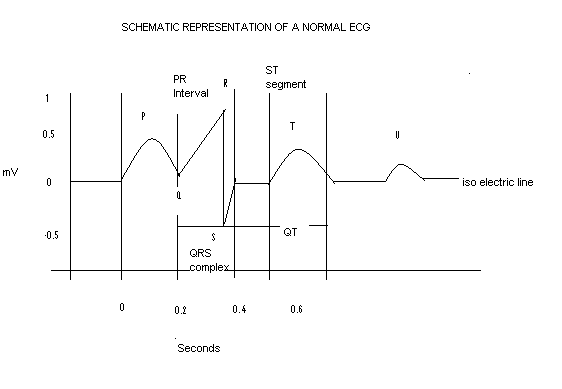
Electrocardiogram (ECG)
This refers to a continuous recording of the electrical activities of the heart. They are obtained by placing sensory electrodes on the surface of the body and recording the voltage difference generated y the heart. “The ECG indicates the overall rhythms of the heart, revealing weaknesses of the different parts of the heart.” (Littmann, 2010) The ECG is recorded by an active or exploring electrode that is connected to an indifferent electrode at zero potential (unipolar recording) or by using two active electrodes (bipolar recording). The echocardiograph amplifies the voltage and causes a pen to deflect in the proportion to the voltage on a paper. The leads (a signal that goes between two electrodes) are of two types; bipolar leads which are commonly referred to as the limb leads and the unipolar leads (V-leads). “The bipolar leads were the first to be developed and were used before the development of the unipolar leads; bipolar leads record the voltage between; the right and the left arm, the right arm and left leg, the left arm and the left leg.” (Pagana, 1998, p.787) The leads can be attached at any point of the limb and the pen makes an upward deflection when the second named point is positive. Unipolar leads are commonly used in clinical diagnosis. “The unipolar leads record the potential difference between an exploring electrode and an indifferent electrode which is a composite pole made of up of signals from other electrodes.” (Littmann, 2010) These consist of six chest leads which are designated V1 to V6.
An electrocardiogram is used to measure the changes in the size of ventricles, aortic diameter and the movement of the ventricular wall (septum) and valves during a cardiac cycle. “The test may use sound waves to create images of the heart and at normal circumstances ultrasound at a frequency of 2.25 MHz emitted from a transducer that also functions as a receiver usually detect the waves.” (Electrocardiogram, 2010)

Description
- P wave- indicates electrical activity associated with contraction of the atrium.
- PR interval- Is the duration between the beginning of activity in the atrium and the ventricle.
- QRS- indicates the onset of contraction of the ventricles.
- ST-segment- Connects the QRS complex to the T wave.
- T wave- indicates electrical activity associated with cells of ventricles.
- Q-T interval- it is measured from the beginning of the QRS complex to the end of the T wave.
- U- wave-Its not always seen and is usually a small wave. (Electrocardiogram, 2010)
ECG is used for the following purposes: Detection and diagnosis of heart conditions (such as cardiac kalemias, myocardial infarctions and enlargement of the heart chamber), monitoring of how well a patient is responding to treatment for heart ailments, and evaluation of patients with cardiovascular diseases.
Measurement of cardiac output using Positron emission tomographic (PET) myocardial perfusion
This measurement is important in assessing ventricular function and determination of myocardial contractile response to medical therapy or surgery. The radioactive tracers used in this procedure include Iodine 131 (131I), Xenon 133 (133Xe) and 131MTC. The radioactive tracers are attached to a carrier that will direct them to the heart and then injected into the patient’s body. A positron camera is used to create an image of the heart on a screen enabling the assessing the condition of the heart.
Chest X-ray and computed tomography
X-ray images are taken using the principle of ionizing radiation to cause the blackening of a photographic film or emulsion. The x-rays are passed through the patient’s chest to the photographic film. “The intensity of blackening caused on the photographic film or emulsion depends on the energy of the incident x-ray.” (Richards, 2010) The image is created on the film because of different energies of the incident x-rays due to interaction with different tissues. This will enable the prediction of a heart condition by examination of the x-ray images.
Computed tomography (CT scan) is a technique whereby multiple x-ray images are combined using a “computer to produce a cross-sectional view.” (Pagana, 1998, p. 779). Cardiac CT scan enables imaging of the “heart with or without using the intravenous contrast dye.” (Pagana, 1998, p. 780) The CT scan may be used to investigate the large vessels, the anatomy of the heart and coronary circulation. “Several types of CT scans are used to detect heart diseases, they include: Coronary CT angiography (CTA), Calcium screening heart scan, dual-source CT and total body scan.” (Pagana, 1998, 780)
- “The calcium CT scan is used in the diagnosis of atherosclerotic plaque in the coronary arteries by detecting calcium deposits in the heart.”(Pagana, 1998) This technique is mostly used in the early detection of coronary calcification due to atherosclerosis.
- “The Coronary CT Angiography (CTA) is a technique that produces high-quality images of the heart and the large vessels” in motion to ascertain whether calcium deposits (plaques) or fatty acids have accumulated in the coronary arteries. Iodine containing contrast dye may be injected IV before the test is performed to improve the contrast of the images produced. (Echocardiography 2008)
- The dual-source is CT provides images from different areas at the same time. Two detectors are used in this case.
- The total body scan entails the production of images of the whole body to ascertain the normal functioning of the heart.
Benefits of Noninvasive Cardiology Technology
Noninvasive cardiology has benefited mankind in many ways which include: “early detection of cardiovascular and heart diseases, reduction of the risks associated with the invasive procedures, reduction of costs, it is highly specific and has gone to a great extent to reduce the cardiologist’s workload.” (Brown, 2010)
Early detection of Cardiovascular and heart diseases
Many heart diseases develop insidiously; According to Littman, it is not easy to tell whether a person is in danger of a certain heart condition by just observing them. (2010)The underlying mechanisms that lead to the different heart conditions have been identified by using non-invasive cardiology technology among other techniques. Patients who are at risk, for example, those with a family history of heart conditions or those with obvious predisposing conditions such as obesity can ascertain the risk of developing heart conditions by undergoing noninvasive cardiology diagnosis.
Fewer risks to the patient
Noninvasive cardiology technology offers the safest way of diagnosing heart conditions. The complications associated with the invasive procedures are very minimal when it comes to the noninvasive option. Although the two types of techniques may be performed together for better results. Noninvasive cardiology technology minimizes the chances for complications such as hemorrhage, injuries and infections due to contamination.
Cost-effective for the patient
Noninvasive cardiology is very cost-effective for the patient considering the time and money a patient will spend to undergo invasive procedures. The non-invasive techniques also allow the early detection of diseases. If a condition is detected early and corrected the patient will pay less compared to treating a fully-fledged disease condition.
High specificity
The results of a medical diagnosis using noninvasive techniques are produced in form of images. The images can be analyzed and a conclusion arrived at with certainty. The results will not be error-prone like in the laboratory tests or the invasive procedures.
Reduction of cardiologist’s Workload
Initially, the cardiologist had to take long procedures to perform an examination or diagnosis. This included the preparation of the patient, creating sterile conditions to operate or carry out invasive procedures, collect for specimens, treat the wounds inflicted and wait for long hours to get feedback. Noninvasive techniques have taken care of this extra baggage. Now, the cardiologists only need to position the patient, set the machines and wait for feedback. This has reduced the cardiologist’s workload to a greater extend.
Challenges
No single good invention comes without challenges. The noninvasive cardiology technique has posed its fair share of challenges. The challenges are not necessary from the technology itself, they include the high initial cost of installation, lack of enough, and adverse effects.
The high initial cost of installation
The cost of acquisition and installation of a functional noninvasive cardiology unit is very high. This has limited the number of users of this technology because it is out of reach for most of them, especially in poverty settings where the problem is compounded by a lack of enough cardiologists and noninvasive cardiology technicians
Lack of enough specialists
The health sector has for a long time grappling with the issue of staff shortage. Noninvasive cardiology is more affected because it is a relatively new field in medicine. The shortage has caused several effects including, scarcity of the service because hospitals lack specialists for them to offer the service. Secondly, it has led to higher costs the specialist available are expensive to maintain, therefore, every hospital that offers the service pushes the cost to the customer.
Adverse effects
The procedures involved in the noninvasive cardiology technology expose the patients to health risks. The use of radionuclides poses a risk of developing cancer both to the patient and the practitioner. The doses offered are designed to minimize this possibility. However, repeated procedures over a short period or errors during the procedure can increase the risk and actually result in cancer development. The same applies to the ionizing radiation used in x-ray procedures. Nevertheless, the benefits of noninvasive cardiology technology out way the adverse effects by far.
Conclusion
Noninvasive cardiology has had a major impact on medicine. The technology has been used to save many lives from threatening heart conditions. It has offered specialists a technique to understand the mechanisms that lead to various heart conditions and therefore a chance to intervene and correct before it is late. The technology was a culmination of many experiments conducted by many researchers. Ironically, the technique was developed by experiments that were supposed to enhance the effectiveness of invasive cardiology. The researchers who developed noninvasive cardiology technology were mainly radiologists who were looking for ways to monitor invasive techniques in the body. The initial techniques were supposed to guide catheters at or close to the heart.
Reference
Brown, E. R. (2010). noninvasive cardiology technology. (writer,Interviewer Echocardiography. (2008). Echocardiography A journal of Cardiovascular Ultrasound and Allied Techniques. Web.
Electrocardiogram. (2010). Web.
Littmann, L. (2010). noninvasive cardiology technology. (Writer Interviewer)
(Electrocardiogram, 2010)Pagana, K. D. (1998). Diagnostic and laboratory tests. Missouri: Mosby.
World health organization(WHO) (2009). Cardiovascular diseases. Web.