Introduction
The Philips MammoDiagnost was a creation of the Philips Medical Systems, and it was brought out in the year 2004. The creator of this system is a part of the Royal Philips. The system has numerous features that are geared towards the increase of mammography safety for both patients and staff. These include features of protection from radiation, measures for ensuring quality assurance, features for safeguarding electromechanical features, and a suitable ergonomic design. The system is designed for safety, reliability and consistency in order to market itself by these qualities (Philips Medical Systems, 2004, 1).
It is interesting to note that the people marketing this product have another additional catchphrase for marketing the product: “Rise to the challenge.” The equipment that is used for specialized X-ray imaging requires radiographers who are highly skilled, and who have the competence to ensure that the procedures of medical imaging are carried out in a safe manner. To utilize the technology used by this product in a pragmatic way, one needs to be aware of the protocols that are used in safely operating this product. The protocol or safe-operation process needs to be written down by the managing radiographer in a manual for safe operation of the machine.
This manual will therefore act as the key document for mammography setting. The manual must also be designed in a user-friendly way to act as a guide to staff for medical imaging that may not be familiar with the system. The manual will also include information on how the machine will be able to adhere to the ALARA principle, i.e. the principle of as low as possible. In order o report on the professional and safe use of the Philips MammoDiagnost, there is need to address four areas.
These include protection from radiation and the safeguarding of electromechanical safety. The latter involves creating awareness among staff about the electrical cables in the machine, and the currents flowing through the cables, which will maximize clinical utilization. It will also make the process of carrying out scans safe and effective, thereby minimizing the necessity of repeat scans. The fourth area to be addressed is the design of a reliable and holistic Quality Assurance program that is designed to be carried out periodically as long as the mammography system operates.
Safe Operation Details
Measures for radiation protection
Radiation protection is perhaps the most vital aspect of radiography, and therefore the need to have a manual for safe operation. The Philips MammoDiagnost, produces superior images and therefore when it is used, repeat scans are rare. This means that patients are exposed to the least amount of radiation possible (Philips Medical Systems, 2004). In mammography, “patient” is used to refer to two possible groups of women. The first group of women is those who have been prescribed to take breast mammographic x-ray examinations. The second group of women is the one that include women participating in programs for breast screening.
Evident in these cases is the fact that the targeted area for the scan is the breast. The same applies to all mammographic procedures. However, in order to visualize and ultimately detect abnormalities in women’s breasts, higher doses of x-rays are used than in most x-rays. It therefore results in a problem among the radiographers who work with mammography. This is because too much increase in the dose of the radiation used in the mammography could make sensitive tissue in the breast induce its breast cancer predispositions (Bushong, 2008). On the other hand, if a low radiation dose is used, the radiographers will be unable to produce an image of the breast that can be effectively used to detect abnormality (Radiological Society of North American, 2009). In cases where this happens, a repeat scan is normally conducted exposing the patient to radiation two times.
The Philips MammoDiagnost (2005) is specially designed to remove this risk and dilemma. The Philips MammoDiagnost is termed as the “optimum dose facility,” and thus the machine has the capability to select the best combination of anode and filter for specific patients. It also has other add-ons such as the specialized system of four focal spots. Its x-ray tube also has a dual-track system. The above features combine to ensure that each breast condition is given a dose that is the lowest possible for it. This is particularly vital for women whose breasts are dense for radiology, especially those who are young (Byng 1998).
There are a number of other crucial radiation protection measures in carrying out mammographic procedures. The forces that are currently in healthcare have a two-faced impact on radiographers. First, physicians are prescribing mammographic scans to more women worldwide due to the increase in the number of aged women all over the world, and due to the quest by physicians to diagnose serious conditions like cancer early. Secondly, radiographers have been given more reporting obligations than in the past. In mammographic procedures, the exposure to radiation is determined by a measure known as entrance skin exposure (ESE).
The ESE is used because of its ease of access with TLDs (thermo luminescent dosimeters). It is now recommended that mammography should use an ESE of 800 mR/view. The potential of the x-ray should not exceed 26kVp because this leads to low image quality that renders the images unusable (Bushong, 2008). Radiographers should also ensure that they are aware of the Glandular dose (Dg) of the patient’s mammography. This is approximately 15% of the ESE of the patient.
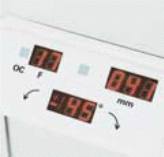
The reason for this is that the exposure of the glandular tissue to radiation can possibly lead to dangerous biological responses. Therefore, an important responsibility of a radiographer is to ensure patients are protected from radiation. With the Philips MammoDiagnost, this is guaranteed due to its optimized compression and its control of radiation dosage that uses W/Rh instead of Mo/Mo (Philips Medical Systems, 2004). The optimized compression is as shown below.
How to safeguard electromechanical safety
Among the first steps in ensuring that mammographic equipment is electromechanically safe is consultation with an electrical utility provider to ensure that the design of the equipment is in conformity with the national and state requirements. In addition to this, the room housing the mammographic equipment must be strategically designed such that the electrical inputs and outputs are properly located to ensure that electrical wires and cords are kept off the ground.
The supply of electricity should be examined in terms of rating, protection (fuses), as well as the provision of earth and isolator. The internal and external wiring of the equipment should be organized. Modifications, extra wiring, and addition of devices should be thoroughly investigated. For the internal wiring, a service engineer has to be present when it is being carried out. Cable protection is paramount especially if the cables cross metal panels and covers. Cables should also be monitored to ensure that they are not strained or stretched. Removal of covers should only be done using tools to prevent possible contact with live parts (Fintor 1995).
Installation of accessorizing equipment should be guided by prescribed instructions and the equipment should be stable. Powered movement in the set-up of the equipment should be operated through ‘dead man’ type controls. Emergency stop devices should also form part of the set-up. Pressure protection should form part of the x-ray tube. Provision of an interlock is vital because it prevents vertical movement and rotation of U-arm assembly by power after compression. The compression device should be designed to apply less than 300 Newton, while the power driven force of compression should be less than 200 Newton. The equipment must be able to release compression after system failure (Fintor 1995)
How to maximize clinical utilization
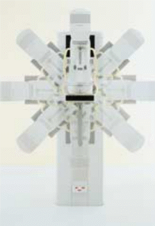
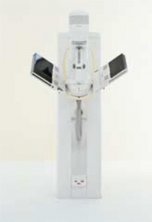
The manufacturers of Philips MammoDiagnost (2005) have designed the machine with many features with an aim of increasing its efficiency and effectiveness. The guarantee of the product is represented as “A versatile system for high volume screening” (Philips Medical Systems, 2004). The system is bound to make a difference is patient traffic after hitting the ground in any radiology department. Below is an image of the system’s “Isocentric Rotation” feature.
In order to increase the utilization of the equipment, the system for patient movement works in two ways. The first way is the placement of the patient in a position of optimal scanning using a motor. This is done as quickly as possible. In addition to this, the system has the capability to maintain accurate height of the object in all possible projections. This two-step functionality of the system ensures that no time is wasted as patients manually position themselves. It also ensures that chances of the occurrence of motion artefacts are decreased.
An incorporated Bucky system and an anti-scatter grid are required in mammographic x-ray equipment in order to determine the quality of the image. Among the advantages of this machine is the fact that it can change collimation automatically by switching between formats. This implies that the machine saves time and allows for more scans.
The Philips MammoDiagnost has other additional features for speeding up its scanning procedures. It has magnification controls that are fast and easy to operate, it gives the radiographer special views and it has a function for stereotactic biopsies.
The design of the machine has also made it more user-friendly, and thus a large art of the population is bound to benefit from it. Bariatric and geriatric patients are also among the people who benefit from the system because of the machine’s maximization of accessibility by patients and its ergonomic design (Philips Medical Systems, 2004). The aforementioned increase in the comfort of users and patients while using the system is attributable to the optimization of dose control and the positioning function of the system.
Quality Assurance
The process of mammography needs rigid Quality Assurance (QA) because of its associated risks. In the first place, unnecessary radiation exceeding the required radiation can cause cancer (Health Canada, 1995). Other scholars believe that if the compression employed by the technique exceeds its limits, tumours can begin to metastasize. That happens in case the patient had a pre-existing tumour. Finally, in order to make good use of mammography, there is need for a high degree of accuracy. A dysfunctional system or wrong parameters will therefore render the technique ineffective. In addition to this, a large number of patients develop emotional trauma after undergoing mammography. It is therefore of essence to ensure that Quality Assurance is of highest degree.
In mammography, it is vital that the equipment is working properly, and that all staff members are conversant with the system in order to benefit the patents fully. A careful analysis of the cost of offering mammography scans will show that expensive resources are needed to start and maintain a credible QA program. A QA program will need well-paid staff, test equipment, and external agencies. In addition to this, test images and staff training add to the cost (Bushong, 2008). Despite these costs, a good QA program has many benefits to the mammography department. The resources will be used well in settings that are well managed. The mammography equipment will also stay longer if the department has a QA program. The flow of patients will increase, and thus the departments will have better funding and better outcomes related to patient care.
A QA in mammography requires a minimum of three team members. In this tem, the radiologist is the leader and thus he/she should be in charge of the other team members. After the radiologist, the next person in charge should be a medical physicist. He/she is the one responsible for the technical issues associated with mammography as well as the physical equipment. The team also needs to have a radiographer. After establishing the team, there is need for a breakdown of annual, quarterly, monthly and daily tasks (Farria 1994). Weekly and daily tasks relate to cleanliness of the mammography room and equipment. Visual aspects of the machine are checked on a monthly basis. Quarterly QA usually involves evaluation of the performance of the mammography in the quarter and the making of necessary adjustments and corrections. For instance, correction repeat scans.
Annual QA involves evaluation of the system as a whole
Indicators
The QA that relates to indicators of the system aims at ascertaining that the system clearly indicates the base status. Additionally, users of the system must be able to access the mains of the system easily, and switch it on or off. The energized status of the x-tube should be clearly shown by a visible sign in the control panel. Users should be able to select either manual or automatic from the control panel.
Collimation and alignment of beam
The safety from radiation should not be compromised by the inclusion of ROI. The medical physicist must check the x-ray to make sure that it is in alignment with the light field. The film and the paddle must be aligned such that the film does not form part of the image.
Radiation output and Exposure Switch
At this level, Quality Assurance can be about the increment of the safety of the patient after each exposure. This level also offers a chance for ensuring that some design elements are practically assessed. For instance, users can ensure that the exposure switch stands 2 meters from the tube focus, and that it has a protective screen.
Half-Value Layer (HVL)
The medical physicist does this section’s QA. It involves the accuracy and reproducibility of kvp. HVL is used in this level to assess the dose rate.
AEC Performance
AEC standing for Automatic Exposure Control is another area where the medical physicist comes in handy during QA. This area is partly involved with the measurement of thickness compensation.
Compression device
Among the most controversial processes of mammography is the process of compressing the breast before the actual scanning. It is therefore vital that this area forms an indispensable part of the QA. The compression, driven by power, should come down to at most 70 N.
System resolution
Image quality and diagnosis are also areas of great concern in mammography. It is therefore vital to acquire quality images for a diagnosis that is based on better information that is more reliable.
Mean glandular dose
The dose to soft tissues like glands must be keenly monitored in order to ensure that excessive radiation does not result. This is because the latter can lead in adverse reactions in the soft tissues.
Summary
To sum it up, mammography is a vital procedure because of its usefulness in early breast cancer diagnosis. The operation of the mammography requires electromagnetic safety, radiation protection, and quality assurance.
For the best service of the mammography system, there is need to reduce the radiation dose, increase the flow of patients, use the available resources more efficiently, increase the budgetary performance of the department and make the mammography equipment last for long. There is also need to have a rigid QA program. This must involve three individuals headed by the radiologist. The others include a mammography specialist and a medical physicist. Tasks are scheduled on a yearly, quarterly, monthly, and daily basis. The radiographer must ensure that reliable QA is put in place because it is vital in radiology.