Introduction
“Hematoxylin and Eosin (H&E) is the most common stain method used in a histopathology lab” (Norman 2009, p.45). Hematoxylin is a solution that stains the cell nucleus to a blue or purple hue. Eosin solution is applied as a counter-stain after (H&E) and acts to stain what hematoxylin did not (Norman 2009). Eosin produces red orange and yellow orange cytoplasm and smooth collagen tissues respectively. For the sake of this practical, there was focus on the human uterus. “This method uses hematoxylin solutions for nuclear staining and eosin solutions for cytoplasm staining” (Cook 2006, p.150). Initially the nuclei are stained with a hematoxylin solution and will stain either blue, dark violet to black. This is then followed by counterstaining with a xanthenes dye for example eosin Y, Eosin B or the erythrosine B. For this kind of process cytoplasm, collagen, keratin and erythrocytes will stain red (Luna 2005, p.401).
With this method of staining, one is able to have an overview of a tissue structure, enabling the determination of structures as being normal, inflamed or degenerative changed or pathological (Eglars 2009). A diagnosis can be made based on the results of this method and is used for the following materials like; paraffin sections, frozen sections and clinical- cytological specimens. They may be uterus, urine, sputum, lavages or effusions. In the long run this method gives outstanding results from the combination, coordinated components, easy to apply as it is already developed, produced and tested (Knowles 2001).
The main principle behind all the hematoxylin process is that the positively charged metal-hematein complexes bind in an acid milieu to the negatively charged phosphate remains of the cell nucleus-DNA. Hematoxylin is then oxidized in the Gill solutions to hematein with sodium iodate as opposed to the mercury oxidation that was used in the past (Raymond 2008). This one is performed in a controlled way, thus more efficient and less harmful to the surrounding.
On the other hand the principle for eosin y which is an acidic colorant that binds mainly to proteins. “The most commonly used is the aqueous eosin y solution in histology” (Walker 2002, p.400). The balancing between nuclear staining with hematoxylin and cytoplasmic staining with eosin is a matter of personal preference. The concentration of the ready-to-use solutions makes it easier for quick obtaining of results (Sudha 2006).
Methodology
A slide was identified from the previous practical session by careful checking of the pots provided to get one that was to be used. A stop watch was used to get the timings right.
All the slides were put together in one holder for the next processes as this was done by a group of students.
“The sections of the specimen were deparaffinized by the two changes of xylene which lasted for 5 minutes each” (Walker 2002, p.36).
The specimen was then rehydrated in 2 changes of absolute alcohol for 3 minutes each. This was continued in 90% alcohol for 3 minutes and then 70% alcohol for 3 minutes.
It was then washed in distilled water briefly and thereafter stained in Giles hematoxylin solution for 6 minutes while the excess was washed in running tap water for few seconds.
The specimen was then differentiated in 1% acid alcohol for 20 seconds and washed in running tap water for 5 minutes. This was followed by counter stain in eosin Y solution for 30 seconds to 1 minute.
The product was then dehydrated through 70% alcohol for 2 minutes, 90% alcohol for 2 minutes and 2 changes of absolute alcohol for 3 minutes each. “After this it was then cleared in 2 changes of xylene for 3 minutes each” ” (Walker 2002, p.37).
“Finally it was mounted with xylene based mounting medium” ” (Walker 2002, p.37).
Light microscopy on the specimen
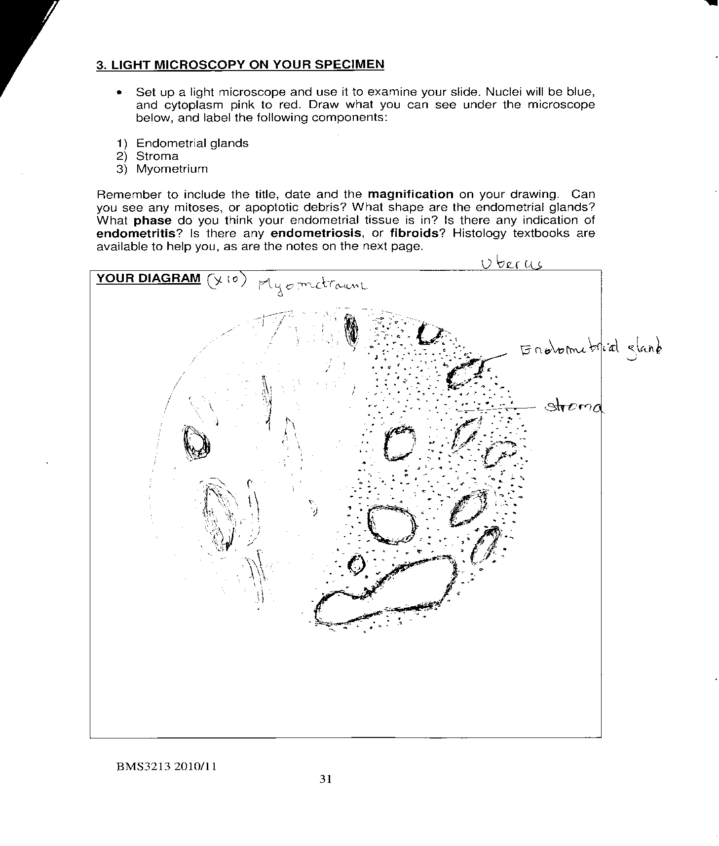
“A light microscope was set up and used to examine the slide. The nuclei were blue, and cytoplasm changed from pink to red” (Jerris & Rick 2009, p.91). The image observed under the microscope was drawn and the endometrial glands, stroma, endometritis and myometrium labeled.
Frozen sections
The specimen was initially frozen rapidly onto a freezing cold metal block with liquid nitrogen and then sliced on a freezing microtome in a special cold cabinet called the crystostat. An anti-roll plate was incorporated to stop the sections from curling up as they were cut, and placed directly on a room temperature slide (Murray 2010).
Results
The following was an image of drawn showing the endometrial glands, stroma, myometrium and endometritis as observed under the microscope with a magnification of x 10.

Discussion
Following the understanding of the hematoxylin and eosin stain behavior on endometrial tissues, the results point out a number of critical issues concerning the composition of the endometrium. In practice, “the endometrium is composed of two layers; the functional layer and the basal layer” (Courtney 2007, p.801). The epithelial layer comprises the stroma with also contains the blood supply. From the theory and results which indicate the blue color for nuclei and red orange cytoplasm and the blood cells. The red orange color seen for the stained specimen can be used to deduce the variations during the menstrual cycle (Hatcher 2008).
The results can also be used to determine the various phases in the endometrium. For instance, there is a phase known as the proliferative phase of endometrium where mitosis is evident in glands that seem to be long and straight in longitudinal section (Buddy 2004). Another phase observed during the experiment was the secretory phase of endometrium where the glands are known to become coiled and complex in form. For the menstrual phase, there are traces of blood seen in stroma. (Raymond 2008).
Hematoxylin and Eosin stain can also be used to test specimen for diagnosis of disease in the endometrium (Anderson 2008). The stain can be effectively used to distinguish inflammations of the endometrium caused by infection and could easily be seen in tissue sections as the presence of acute inflammatory cells in the endometrial glands. This often occurs after childbirth, and usually more than one organism can be cultured (Bancroft 2002).
Conclusion
Hematoxylin and eosin stain is thus quite essential for experiments and laboratory tests involving a number of tissues to give the necessary diagnosis. This explains why the stain has found a wide application for many years. The stain, as seen from the experiment helps in identifying various types of tissues and changes in morphology. H&E stain works both as a stain and counter- stain to display a wide range of features in the nucleus, cytoplasm and the cell in general. With this method of staining, it was possible to have an overview of a tissue structure, enabling the determination of structures as being normal, inflamed, degenerative changed or pathological.
Having successfully performed the experiment to achieve the desired result, the practical thus met its objectives. The application of hematoxylin and eosin stain as well as frozen section preparation was well appreciated.
References
Anderson, A., 2008. Principles of regenerative medicine. California: Harvard university press.
Bancroft, D., 2002. Theory and practice of historical pathology. Michigan: Wiley and sons publisher.
Buddy, D., 2004. Biomaterials science: an introduction to materials in medicine. Washington: Academic press.
Cook, J., 2006. Cellular pathology: introduction to technique and applications. New York: Scion Publishing limited.
Courtney, M., 2007. Elsevier Medical images: hematology and eosin staining. New York: McGraw publishers.
Eglars, A., 2009. Bone & soft tissue pathology: a volume in the foundations in diagnostic pathology series. Colorado: Saunders press, 2009.
Hatcher, M., 2007. Advanced techniques in diagnostic cellular pathology. New York: Wiley&son’s publisher.
Jerris, R. & Rick, E., 2009. Clinical procedures in emergency medicine: expert consult- online and print. Colarado: Saunders.
Knowles, D., 2001. Neoplastic hematopathology. London: Oxford university press.
Luna, G., 2005. Histological staining methods of the armed forces institute of pathology. New York: Mc Graw hill.
Murray, L., 2010. Oxford handbook of clinical medicine. Oxford: Oxford university press.
Norman, F., 2009. Ultra structural pathology: the comparative cellular basis of disease. Chicago: Wiley & sons publishers.
Raymond, R., 2008. Cell & tissue based molecular pathology: a volume in the foundations in diagnostic pathology series. Minnesota: Churchill livingstone.
Sudha, R., 2006. Thyroid cytopathology: an atlas and text. Cambridge: Harvard university press.
Walker, J., 2002. The protein protocols handbook. Washington: Harvard university press.