Introduction
Tuberculosis is among the disease category of rare bone diseases, and the problem is estimated to occur in the range of 1 to 3 %. Despite the fact that the disease is termed as rare, its occurrences are increased with the provision of certain favorable environments/conditions such as malnutrition, drug, and medical abuses, presence of certain diseases and/or conditions like HIV, diabetes as we as immune-compromising factors among others. In addition, bone tuberculosis is usually noticed as lesions occurring singly, but the disease sometimes presents with multiple lesions in different patients. Whereas certain diseases show specificity in the area or parts of the attack, bone tuberculosis appears to be least specific, and thus universally infects any type of bone regardless of size and position. The spinal bones are most likely to be infected while attacks are less commonly found in long bones, as it is according to Madkour and Warrell’s (1) knowledge and understanding of the nature of the disease.
It is also noted that the problem is likely to originate in parts that are more vascularized in nature. This is evidenced by its common origination at metaphyses in children. Moreover, tuberculosis cases though are noticed in all ranges of the demographic population of people, young children and adults depict high prevalence of all tuberculosis forms. In this view, Golden and Vikram (2) believe the above generalization of the disease’s ability to attack the various body part (tissues and bones) is a clear indication and suggestive of how the disease might present itself as a global threat.
Diagnosis
There are various diagnosis methods available for detection of the bone tuberculosis. The diagnosis of the bone tuberculosis may be done through the employment of imaging techniques, laboratory probing and/or utilization of clinical methods. However, the employment of the laboratory technique is generally not encouraged for results do not count much in the diagnosis and intervention process due to their high level of unreliability.
Imaging method/ techniques
Bone tuberculosis can be detected and diagnosed by the use imaging techniques. This implies the methods are much more reliable during establishment of the disease problem together with the subsequent intervention process. Fausto et al (3) suggests some of the most commonly used imaging techniques for the bone tuberculosis include magnetic resonance imaging, which is plays crucial role in the detection of bone marrow edema during the initial bone attack by the disease. Computerized tomography- used in the detections of vascular malfunctioning and problems such ischemia and vascular congestions. The last method is use of plain radiography, which is normally employed when the bone disease activities are already eminent and clear.

X-ray radiography
During the diagnosis of bone tuberculosis, radiologists require to examine various parts of the bone area. This means that depending on the problem description by the person seeking medical help, radiologist may perform a single radiographic view or several of them. However, Madkour and Warrell (1) warns individuals on the risk associated with radiographic processes and further wisely advises that it’s better to carry a single radiographic investigation to minimize the risks associated with radiation effects. Generally, a radiologist may investigate the disease problem through carrying out of a frontal projection or lateral view.
Example in elbow TB
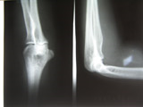
Goroll and Mulley (4) describes that in case a client express an enlarging joint accompanied with restriction on movement, the radiologists take a lateral radiograph at the elbow point of the specific hand but no any other part of the body. The isolation of the hand from all other parts is a measure taken to ensure that the X-ray radiation does not fall on any other body part. This prevents causing damage to the cells and organs that lies outside area of target. The part is also specifically selected because it has the high possibility of being attacked by the disease problem. The idea is that the investigation is localized and therefore the technique helps to save time during the process of probing
The adoption of these radiographic techniques therefore has several advantages and benefits in the carrying out routinely diagnostic procedures. The method helps radiologists to obtain immediate relevant information of the anatomical structure of a mal-positioned or misaligned part of the body. They give structural details and information of both the tissues, cells as well as the bone structure during radiologic analysis which be used by nurses and other persons to gain deeper knowledge and understanding of the anatomical parts which they might have not studied before. Thus, there is possibility of discovering new research topics, or even resolving structural/anatomical problems that have been contradictory over the years. Under the proper use of the technique, Deshmukh et al, (5) states that the patient’s risks are quite minimal as nothing is gained nor lost from the patient.

Above is X-ray Images showing lateral and front radiographs of the elbow.
Conclusion
Finally, the method is seen as of great advantage as it quicker in diagnosis process, and ensures precision besides permitting the checking a diversified number of disease problems, that is, during the diagnosis of tuberculosis, the medical practitioner can also determine problems of edema and diabetes.
References
Madkour M. and Warrell D. Tuberculosis. New York: John Wiley & Sons; 2006.
Golden, M., and Vikram, R. Extra-pulmonary tuberculosis: an overview. American Farm Physician; 2005. 72:1763–7.
Fausto N., Mitchell, N. and Vinay K., Robbins Basic Pathology. Philadelphia: Saunders Elsevier; 2007.
Goroll A. and Mulley G. Primary Care Medicine: Office Evaluation and Management of the Adult Patient. Lippincott Williams & Wilkins; 2009.
Deshmukh T., Hoskote S., Iyer R., and Sanghvi A. MRI features of tuberculosis of the knee. Journal of Skeletal Radiology; 2009. 38:269–74.