Abstract
Evident from previous research and casual empiricism, application of 3D ultrasound technology across many healthcare centers demonstrate much promise. This shift in philosophy has forced researchers to keep an eye on current technological changes. 3D ultrasound application in the modern medical world is inevitable and may be considered to possess both positive and negative impacts. The methodology in this research utilizes 3D technology of capturing multiple-dimension images on pregnant women while focusing on the progressive development of the fetus in its entire gestation.
Introduction
Technology has improved the way medical specialists diagnose diseases. The newly introduced technology such as 3D ultrasound has increased the likelihood that tissue abnormities can be detected with no much effort. Evidently, modern 3D surfaces have demonstrated clinical utility. Ultrasound is an emerging modality and research argues that its application to have improved diagnosis in human organ soft tissues that would have otherwise missed by standards MRI procedures. An extrapolation from 2D ultrasound technology, 3D imaging has undoubtedly increased the evaluation of standardization of ultrasound evaluation. As 3D ultrasound continues to mature, this research will investigate specific 3D ultrasound application to clinical set ups.
Evidence-based practice is the basis of work for most practitioners, in which the same evidence is used but is being applied for different purposes. 3D modeling technology has provided avenues for a variety of technological development, especially in the medical field. The technology has been embraced by a wide variety of ultrasound laboratories as a useful diagnostic tool. Widely applied in management of pregnancy, 3D technology non-invasive procedures have made is easy for parents to know the sex of the baby as well as rule out any anomalies. 3D models imaging technology works by transferring media images into health and diagnostic procedures enabling parents view video of their child prior to delivery.
This study is aimed at identifying both positive and negative influences of computer technologies as important diagnostic tool. The methodology is based on secondary data, statistics were provided as well as primary data in form of a questionnaire that will be given out to several participants in order to form a qualitative empirical analysis on this topic. The survey will be measured in a Likert scale that will enable patients’ rate their opinions regarding use and application of 3D ultrasound technology
After gathering the replied surveys, the aim of the study is to compare and contrast the results between hybrid pregnant women and real expectant mothers as well as with the literature review in order to make clear to the reader how innovative technologies in the modern world, how it is changing with evolving healthcare and finally examine if such applications have positively influenced activities of sonographers.
Literature Review
Applied as a medical ultrasound technique, three-dimension (3D) imaging is used as a diagnostic tool in determining fetal development at various stages. It provides a three dimensional image that shows features of a fetus including movement and the sex of the baby. Ultrasound technology has for the past years experienced dramatic improvement as quoted by Lazebnik and Desser (2007) in “ultrasound image quality, resolution, availability and a range of indications” (p.1). Evidence-based practice is the basis of work for most practitioners, in which the same evidence is used but is being applied for different purposes. This is coherent to the 3D planar imaging are improved technology previously applied in the 2D ultrasound technology. The essay will record experiences in research (i.e., approach, methodology, critical thinking) may be considered evidence of practice in themselves since the concepts about how they are done are being applied practically. These advances accelerated the explosion of various medical applications whetting appetite for more advanced volumetric CT and MRI technologies. The rigidity of tasks undertaken in the previous 2D imaging stimulated sonographer’s appetite to provide a comparable volumetric technology that is more advanced.
Physics of 3D Ultrasound
As an extrapolation from 3D technology, 3D ultrasound is applied as a medical diagnostic technique that utilizes waves within the ultrasound machines. The beams reflected from the scanned image are transmitted back to the transducer projecting an image onto the screen in form of a video. Different scanning models exist in obstetric ultrasound, but 3D ultrasound features are specifically designed to pick sound waves being sent to different angles. Sound waves sent back and forth return with echoes which are processed by a computer program projecting in three dimensional images of internal organs. 3D ultrasound uses the same technology but clearly defines width, heights and depth of images for proper diagnostic analysis.
Created by Olaf von Ramm and Stephen Smith at Duke University in 1987, it was though that 3D ultrasound imaging technology would ease clinical diagnosis especially in fetal anomaly scanning, breast and pelvic imaging. Deeply embedded in 3D technology, picture display in 3D ultrasound is completely real time and eliminates lag problems associated with delayed computer constructed images common in traditional MRI ultrasounds.
Benefits and Limitations of the 3D ultrasound Technology
It is indicated that radiologists dependent on a series of non-continuous and often uniform imaging organ as a prediction of the whole human anatomy. This therefore requires repeat examination of sampling in developmental which is often challenging and time consuming since fixing of the imaging plane and reconstruction of other cross section are required. Second limitation is that no spatial relationship is indicated in the images submitted for review, forcing a radiologist to depend on image labels trusting the labels have minimal sonographer variability. To verify this, a sonographer is forced to perform a comparison analysis for the acquisition technique used, which proves challenging in an event where serial exams were performed since no exact corresponding planes will be acquired. Third, volume measurements such as ellipsoid are defined by length and width and do not depict the true 3D shape. Finally, transformation from 2D to 3D technology does not exploit volume rendering techniques.
Therefore, 3D ultrasound approaches were developed to allow the acquisition of a sonographic volume. It also argued that personal experience of the sonographer in the 2D post-processing cine clips study is also one that may be categorized to be similar to offline workstation acquired by freehand scanning into 3D volumes. Such post-processing scanning are what Lazebnik and Desser (2007) categorizes as “easy to implement and less accurate than volumetric acquisition” (p.1). The 3D technology intensifies data into a 2D image to obtain a true 3D sonographic volume then uses these rays to visualize 3D ultrasound data. In This case, options such as 2D transducer arrays, mechanical scanners and freehand techniques with automated localization are utilized to acquire acquiring sensors of a convectional 2D diagnostic ultrasound machine. This is an advantage because the technique does not involve use of ionizing radiation since it acquires volume information directly a series of continuous or non-continuous 2D images which retains scanning flexibility. Being an extrapolation from 2D technology, 3D ultrasound o acquires sensors of a convectional 2D diagnostic ultrasound machine.
Another benefit of 3D ultrasound is that its sonographic features are universally applicable. First, in 3D ultrasound setting, a sonographer scans the region of interest with a linear-array transducer contrary to the CT scan that requires multiple sweeps. Also, in applications such as bedside neonatal 3D neoro-sonography, 3D ultrasound has greatly reduced time performed on scanning since it eliminates sedation techniques such as the ones performed in MRI modalities (Fritz et al, 2005, p.299). Similar to volumetric CT data, 3D ultrasound volumes are performed prior to the set acquisition in order to rule out any technical problems that may be encountered during a scan analysis. Pretorius and his colleagues (2005) also state that “ an evaluation of multindodular thyroid glands or fibroid uteri, for example, may become much easier when the organ is viewed in multiple planes simultaneously on tomographic display workstations” (p.941). In this sense, Pretorious et al (2009) provides that “post-processing allows remote interpretation and teleradiology for sonography, as the reader has necessary information in the scanned volume” (p.942). This enables easy comparison of serial imaging over a period of time since numerous corresponding anatomic landmarks are displayed in all data sets. Conclusively, this experimental learning is experienced by the sonographer in his engagement with the various learning tasks. Lazebnik and Desser (2007) add that measurement of organ volumes with 3D surface visualization are directly computed by manual/ automatic segmentation that comes along with data set-which does not require interpretation of any specific geometry to analyze scanned images.
Abdomen and Pelvis Application
3D ultrasound benefits are numerous. For example, examining the abdomen with 3D ultrasound requires what Lazebnik and Desser (2007) quote as “estimating volumes of liver masses, gallbladder, or gallstones and other size measurements i.e. kidney long axis, have traditionally relied on the sonographer’s accurate imaging of this plane” (p.3).
Accessibility and Feasibility of 3D Ultrarsound
3D ultrasound already demonstrates clinical utility as evidenced from obstertric applications mentioned throughout this essay. Given that several limitations persist in 2D imaging, the unmatched benefits of 3D imaging are overwhelming. 3D ultrasound also show potential benefits in obstetric imaging (Benacerraf et al, 2005) and when exploring gynecological applications, 3D imaging is regarded as a more efficient clinical approach. 3D is also beneficial in what Benacerraf et al (2005) states as “evaluation of congenital uterine anomalies, where post-processing into the coronal plane permits visualization of the uterine fundal contour” (p.1249). Images obtained here can be used in comparing two volumetric techniques for estimating the nature of fibroid uterus growth. Lazebnik and Desser (2007) also mention 3D imaging application services to include “endometrial polyps, corneal ectopic pregnancies, intrauterine devices, and adnexal lesions and other interventions including abscess drainage of the pelvis and abdomen as well as fertility procedure” (p.3). They mention these medical interventions to have been made easier by 3D technology by enabling viewing of multiple planes at once. 3D imaging also offers benefits in urologic sonography when applied in both urodynamic imaging and anatomic survey. In pediatric application, Mitterberger et al (2006) quotes “3D ultrasound is intrinsically superior to 2D in documentary congenital renal anomalies and ureter configuration (in context of reflux), as these 3D structures cannot be completely visualized in a single plane” (p.11). When further comparing with 2D sonography, Mitterberger et al (2006) also add that 3D ultrasound is superior for evaluation of hematuria with regard to identifying bladder cancer, bladder wall hypertrophy, bladder diverticula, mucosal bladder folds, and re-growth of the prostate, as validated by cystoscopy and/or bladder biopsy” (p.11).
Lazebnik and Desser (2007) mentions that 3D ultrasound imaging in the scrotum provides better 3D visualization of the AUS complex geometry of the epididymis and other extratesticular structures, substantially improving diagnostic confidence and proper medication. For patients with suspected prostate cancer,
Sauvain at el (2007) mentions 3D application in Doppler sonography to “improve diagnostic and staging accuracy of anatomic imaging through improved depiction of prostate vascular structures” (p.30). Finally, utilization of 3D imaging in biopsy site selection has helped greatly in identifying areas of abnormal blood flow. Boggers et al (2008) concludes by adding “extracapsular involvement is evaluated by detecting the presence of vessels perforating capsule and use of microbubble-based contrast agents further increases the sensitivity of 3D ultrasound for prostate malignancy” (p.97).
Breast Imaging
In breast imaging, Cho et al (2006) embraces the 3D technique in its ability to identify breast masses. In stressing their point, Cho et al (2006) state “multiplanar capability of 3D has introduced to the imaging palette the coronal plane, which some investigators suggest improves depiction of tumor margins and of the orientation of tumor relative to ductal structures” (p.31).
Cardiovascular Application
In cardiovascular application, 3D ultrasound has proven beneficial in vascular imaging by providing detailed anatomic structures that includes color, spectral visualization and quantification of blood flow. On the contrary though, Kripfgans and his colleagues (2006) mention the inherent 2D ultrasound contradictions to include inaccurate estimation of flow volume since cross-sections areas are unknown. Recent research by Kripfgans and his colleagues (2006) suggests that “3D techniques provide true volumetric flow estimates that are angle-independent and allows direct visualization and quantification of plaque volume unlike 2D ultrasound that allow detection of arterial atherosclerosis primarily through velocity measurements” (p.1305). AbuRahma et al (2007) further add “the addition of 3D color toDoppler information may provide benefits in the grading of stenotic lesions with respect to flow dynamic” (293). Busek et al (2005) and Klotzsch et al (2008) argue that the ability to reformat 2D volumetric data to fit 3 dimensions enable greater visualization of data flow in a plane resulting to low interobserver. Also 3D provides a single plane that displays all the related vasculature (for example, the hepatic arterial system) enabling the viewer to view the entire anatomy. the viewer can observe the entire tree in 3D. Since the heart’s contraction motion is depicted in a three-dimensional image, the evaluation of cardiac anatomy alongside its function through 3D technology provides accurate measurement of chamber volume and ejection fraction. Ota et al (2007) summarizes that “while 2D echocardiography requires assumption of a simplified geometric mode of the ventricular shape, the 3D approach allows for direct ventricle segmentation” (p.93). They conclude by adding that visualization of an entire valve has been made possible by 3D technology by improving septal defects and analysis of anatomic relationships in congenital heart disease (Houck et al, 2006, p.1092).
Impacts of 3D ultrasound to Professionals
3D ultrasound has been beneficial in real time visualization by guiding of both invasive and operative interventional procedures. Application of 3D ultrasound to neurosurgical procedures has greatly benefited surgical specialists in cases of resection of intracranial tumors. Lindner (2006) provides that 3D information “allows surgeon to modify preoperative planning maps to account for warping and tissue removal” (p.1975) as the applied Doppler imaging provides improved visualization of vasculature of interest. Unsgaard et al (2006) and Woydt et al (2005) further mentions 3D beneficial features to include “biopsy guidance, resection guidance, arteriovenous malformation localization (and involved in vessel identification, localization of peripheral ancurysms, and declination of cavernous hemangiomas in both brain parenchyma and the brain stem” (p.235, 28). For gliomas resection, Unsgaard et al (2005) mentions that “3D ultrasound volumes provide delineation of metastases and solid component at least as reliability as navigated 3D MRI” (p.1269). In summary, 3D ultrasounds does no require data pre-set unlike the MRI application that may be affected navigational accuracy.
Materials and Methods
The methods utilized for analyzing research question were survey questionnaire distributed to each e pregnant woman participating in the research study. The questionnaires were given a study format with a thorough explanation of what was required from them. This strategy was used in anticipation that extensive data will be collected on how each member perceived changes and developments of their fetus rather than forcing themselves into YES and NO replies on pre-existing scales designed around the authors beliefs. Questionnaire method of data collection applied by the research assistants to record raw information and report the findings on the sensitive issues of information technology was reliable and improved the credibility of data collected. In determining the validity the results obtained, generated 3D hybrid models were compared with real expectant mothers with the supervision of obstetricians and radiologists.
Lazebnik and Desser (2007) intensive research into 3D ultrasound technology confirmed that “the study of interactions between radio frequency waves and biological tissues requires precise models of the human body at various stages” (p.1). To this, the research identifies a hybrid human head models based on magnetic resonance image (MRI) data, as a sample for studying impacts of 3D technology to pregnant mothers. Participants were informed on the procedure to be followed and professionals were exposure magnetic fields prior to the research to check if the machines were set correctly. Lazebnik and Desser (2007) define dosimetry as “numerical simulations computing the dose absorbed by body tissues resulting from the exposure to some specified radiations” (p.1). Dosimetry will therefore use a standard method of anatomical model to calculate the absorbed radiations by pregnant women. To do this, detailed information of the tissue including tissue localization, anatomic details of the utero-fatal unit and the organ shapes will be required. The hybrid models of pregnant women were pre-selected on the basis of dosimetry simulations.
Sample
The samples consisted of 15 participant and only 13 responded were responded were assumed to have used 3D ultrasound at least once a month. Three hybrid pregnant women were also used. The response rate was rated at 70% since 10 participants applied 3D ultrasound in one way or another. The responded included a 23 woman, a secretary at department store and a 30 year old housewife woman. The questions were carefully drawn to avoid misrepresentation and where technical terms were used, were followed by simple explanations to ensure higher response rate.
Likert scale enabled participants to indicate their degree of agreement with the statement using a five point scale that indicates;
- Strongly disagree
- Disagree
- Neither agree nor disagree
- Agree
- Strongly agree
Results
3 generic hybrid models series were created to represent pregnant women at a different gestation stages and placed the fetus in different positions. For proper dosimetry scanning, the models were acquired during regular clinical follow-ups and detailed tissues of the fetus and the utero-fetal unit, were displayed. Adequate time was allocated to validate the accuracy of the 3D ultrasound machine with the approval of clinical obstetricians and radiologists.
A total of 13 respondents were used for the analysis after filling and returning the questionnaire for their completeness and consistency. The participant profiles were presented per the following demographic criteria
- 58% of the corresponds were aged more than 23 years
- 59% of the respondents had stable jobs and their able to afford 3D ultrasound
- 24.8% of the responded said heathcare did not cover 3D, so they could not afford.
The age ranged from 20 to 35 years
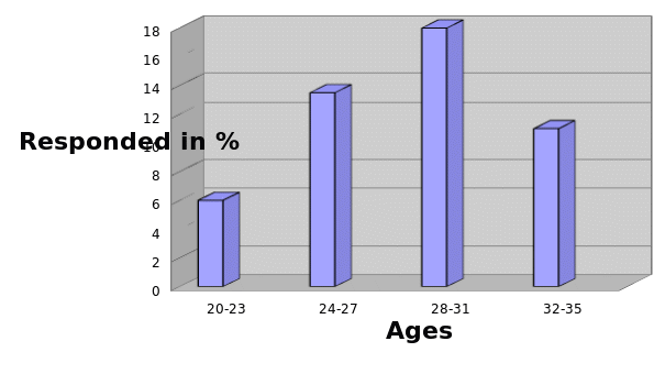
The maximum respondents lied between ages 25 through 30 years whereas minimum respondents were represented by ages 31 through 35. With regards to employment status, only 26.8% of the respondents were permanently employed while the minimum groups of 6% were either jobless or were housewives. The frequency use of 3D ultrasound was measured in monthly basis, and 26.8% argued the technology improved the bonding with child.
Limitation of 3D ultrasound
First; as sonographers would make us believe, 3D image acquisition is not different from the 2D ultrasound since they both use the same contrast-acoustic impedances. Also in obstetrics applications, 3D renderings have been effective in identifying amniotic fluids since they provide high contrasts between the background and the fetal surface but fails to provide the same contrasts in other organ systems. Secondly; 3D platforms does not provide standard display convection making it complex and hard to master or even interpret the reconstructed images. Also, the scientific characterization that uses medical techniques such as posterior enhancement is difficult to understand in an event where multiple simultaneous transducer orientation is applied. Third; 3D ultrasound technology impresses the methodology that does not use present data acquisition, which is a standard methodology employed by 2D for easy interpretation. Fourth; Lazebnik and Desser (2007) quote “rendering of data in 3D using volume or surface rendering techniques introduces an additional layer of potential artifacts and lack of standardized review” (p.4). Fifth; since 3D images are more accurate that the traditional 2D sections, literature that support clinical benefits of 3D ultrasound imaging are insufficient, particularly in comparing their benefits and indications.
Elective 3D ultrasound
Elective 3D ultrasound refers to what Michailidis and his colleagues (2006) states as “ultrasounds performed on pregnant women for the reason of a woman wanting to see her unborn baby including the sex of the child” (p.215). Although there is no conclusive evidence to show the benefits of 3D ultrasound to a mother and the unborn baby, medical literature mentions 3D rays to be harmful when the duration of ultrasound exposure, intensity of the waves and frequency of ultrasound the sessions are uncontrolled.
Duration
Incases of malfunction, As Michailidis et al (2006) states that “3D ultrasound machines should be preset to send signals warning or shut off in cases of breakdown if any of the built-in barriers fail to control the limit of the ultrasound waves as provided by the FDA standard” (p.216). As Michailidis and his colleagues (2006) succinctly concludes, “a higher intensity of ultrasound waves are used to detect the baby’s heartbeat, and as the waves are directed and focused onto a single organ in the fetus, it is advised to use ultrasound machine to detect and play baby’s heartbeat after two weeks of gestation” (p.216).
Frequency
It is however advised use ultrasound at least once a month since studies claim that frequency use may pose potential risk to a patient and should not be substituted with routine prenatal care. Michailidis et al (2006) extensive research also provide that neither of the participants (mother and the child) can feel the heat produced by the machine however higher intensities create slight warmth and may be dangerous to the baby.
Medical effects
There are currently no found reports on the mental defects of ultrasound usage to both the mother and the child.
Other risks
Some potential risks inherent in 3D ultrasound technology are miscalculations that may display false positives which can cause panic to the mother. Inexperienced technician can also misdiagnose certain conditions by using 3D ultrasound machine since some artifacts can be easily translated as duplicates or even missing. It is also advised not to perform ultrasound on fetus less than 17 weeks of gestation as the waves may cause harm to the mother.
For risk reduction, FDA requires that qualified ultrasound technologists with ARDMS-certificates allowed in performing ultrasound scans. Since there is no law requiring 3D ultrasound to be conducted by certified ultrasound technologists, healthcare centers should make an effort to employ qualified medical directors and provide the needed training to perform ultrasound scans. Pregnant mothers on the other hand to produce pre-natal care before 3D ultrasounds are performed. For clear images patients may be advised to drink plenty of water prior to the appointment for adequate amniotic.
Future Developments and Conclusion
Benefits of 3D ultrasound to pregnant mothers are overwhelming, and to this we recommend that 3D ultrasound to be part of routine care, and prenatal clinics should provide them as courtesy. Medical researchers are constantly faced with the task of innovating new technological tools but insurance companies’ serves as a barrier to this, especially if it delivers predictable returns in the short term. In this case, insurance companies should start accepting 3D ultrasound as part of health cover. Medical companies should invest in creating awareness on benefits of elective 3D ultrasounds as it has proven very useful in detecting fetal anomalies which can help diagnosis.
Since the application of innovation technologies relies heavily on medical practices, the selection of suitable computer programs and qualified technological sonographers is required to be able to cope with the technological changes as success or failure of a diagnosis relies on them. This study should also be conducted in several hospitals to compare the results and use large samples from different
States to improve results achieved. The study covered the application of 3D ultrasound innovative technology in the modern healthcare and even considered elective 3D imaging a effective in early anomaly detections; hence researchers may consider studying reasonable limits to which pregnant women should be allowed to take 3d Ultrasounds. Insurance companies should therefore welcome this technology by including it in medical covers.
Evidence-based practice is the basis of work for most practitioners, in which the same evidence is used but is being applied for different purposes. In this regard, research in improving the current technologies should be embraced. 3D modeling technology has provided avenues for a variety of technological development, especially in the medical field. The technology has been embraced by a wide variety of ultrasound laboratories as a useful diagnostic tool. Widely applied in management of pregnancy, 3D technology non-invasive procedures have made is easy for parents to know the sex of the baby as well as rule out any anomalies. 3D models imaging technology works by transferring media images into health and diagnostic procedures enabling parents view video of their child prior to delivery. Since 3D images are more accurate that the traditional 2D sections, literature that s clinical benefits of 3D ultrasound imaging should be provided, particularly in comparing tits benefits and indications from other scanning machines. Although there is no conclusive evidence that shows benefits of 3D ultrasound to a mother and the unborn baby, duration of ultrasound exposure should be limited to only once a month.
References
AbuRahma, A.F., Jarrett, K.,& Hayes, D.J., (2007). Clinical implications of power Doppler three-dimensional ultrasonography. Vascular, 12(5), 293-300.
Benacerraf, B.R., Benson, C.B., Abuhamad, A. (2005). Three- and 4-dimensional ultrasound in obstetrics and gynecology: proceedings of the american institute of ultrasound in medicine consensus conference. Journal of Ultrasound Med, 24(12), 1587-1597.
Bogers, H.A., Sedelaar, J.P., Beerlage, H.P (2008). Contrast enhanced three-dimensional power Doppler angiography of the human prostate: correlation with biopsy outcome. Urology, 54(1):97-104.
Bucek, R.A., Reiter, M., Dirisamer, A. (2005). Three-dimensional color Doppler sonography in carotid artery stenosis. AJNR, 24(7), 1294-1299.
Cho. N., Moon, W.K., & Cha, J.H (2006). Differentiating benign from malignant solid breast masses: comparison of two-dimensional and three-dimensional US. Radiology, 240(1), 26-32.
Fritz, G., Riccabona, M., & Weitzer, C. (2005). Three-Dimensional ultrasound (3DUS) of the neonatal brain: clinical application in patients of the neonatal intensive care unit (NICU). Ultraschall Med, 26, 299-306.
Houck, R. C., Cooke, J.E., & Gill, E. A. (2006). Live 3D echocardiography: a replacement for traditional 2D echocardiography? AJR, 187(4), 1092-1106.
Klotzsch, C., Bozzato, A., Lammers, G. (2008). Contrast-enhanced three-dimensional transcranial color-coded sonography of intracranial stenoses. AJNR, 23(2), 208-212.
Kripfgans, O.D., Rubin, J. M.,& Hall, A.L. (2006). Measurement of volumetric flow. J Ultrasound Med, 5(10), 1305-1311.
Lazebnik, R.S., & Desser, T. S. (2007). Clinical 3D ultrasound imaging: beyong obstetrical application. Continuing Media Education, 1, 1-6
Lindner, D., Trantakis, C., Renner, C. (2006). Application of intraoperative 3D ultrasound during navigated tumor resection. Minim Invasive Neurosurg, (4):197-202.
Michailidis, G.D., Papageorgiou, P.,& Economides, D. L. (2006). Assessment of fetal anatomy in the first trimester using two- and three-dimensional ultrasound. The British journal of radiology, 75 (891), 215–219.
Mitterberger, M., Pinggera, G. M., Neuwirt, H. (2006). Three-dimensional ultrasonography of the urinary bladder: preliminary experience of assessment in patients with haematuria. BJU, 11, 1.
Ota, T, Kisslo, J., Ramm, O.T., & Yoshikawa, J. (2007). Real-time, volumetric echocardiography: usefulness of volumetric scanning for the assessment of cardiac volume and function. J Cardiol, 1, 37.
Pretorius, D.H, Lev-Toaff, A. Nelson, T.R., (2005). Feasibility of performing a virtual patient examination using three-dimensional ultrasonographic data acquired at remote locations. J Ultrasound Med, 20(9), 941-952.
Sauvain, J.L., Palascak. P., Bourscheid, D. (2007). Value of power doppler and 3D vascular sonography as a method for diagnosis and staging of prostate cancer. Urol, 44(1), 21-30.
Unsgaard, G., Rygh, O. M., & Selbekk, T. (2006). Intra-operative 3D ultrasound in neurosurgery. Acta Neurochir (Wien), 148(3), 235-253.
Woydt, M., Horowski, A., & Krauss, J. (2005). Three-dimensional intraoperative ultrasound of vascular malformations and supratentorial tumors. J Neuroimaging, 12(1), 28-34.