Introduction
Ultrasound is sound energy in the form of wave which has increased above 20 KHZ (Srbely 2009, 3). Generally, the human beings can only detect a maximum of sound frequency not more than 20,000 Hz. So far there are only two vital types of Ultrasound diagnostic and therapeutic that is well known. In point of fact, Medical imaging provides the most perfect task of diagnostic to Ultrasound, whereas, the main usage of therapeutic Ultrasound is to treat the numerous types of diseases and disorders in human beings (Srbely 2009, 3).
Towards the turn of a new decade, there has been various technological advancement in machinery of ultrasound; in fact, the new improvements have provided more reliable and accurate ways of treating all diseases and disorders treatable under these category and the outcome of these developments are seemingly more advantageous since they save time and the patient incur less pain compared to the past (Nicholas 2010). Therefore, in this paper we shall assess the two major technological developments in real time imaging: extended file of view and 3-D Ultrasound. Moreover, this paper will discuss the numerous benefits that have been brought about by both advances in real time imaging for the conventional imaging.
Extended field -of -view in Ultrasound
The first chief developments is the Extended field of view (EFOV) which urbanized shortly after the production of static image by transmitting the 1-D probe through the assessing the region utilizing “mechanical positing arm” (Poon and Rohling 2005). However, the process of producing the static image (D-2) was slow due to complication and it has not been included in the real time. For this reason, new developments in Ultrasound have led to using real time imaging. The extended view approach is based on “sweeping” the probe in sideways direction on the targeted section (Poon and Rohling 2005).
The advanced approach -Extended field- of- view in Ultrasound can provide high quality panoramic image even without the need to change the resolution when being used manually. Additionally, the mechanism of Extended field-of-View technology is more advanced based on the fact that it can be used to evaluating of the probe by comparing the sequential images produced through the different types of motion of the probe. Such images are transmuted geometrically accordingly in a careful manner to ensure quality result is established, the images are then inserted in (the Extended filed-of-View) image buffer and later integrated with the other images to provide a quality EFOV (Extended field-of-View) image (Kim, et al. 2003). An EFOV image is more advanced and can also enable recording of images measuring up-to to 60cm long raised above the ground. Furthermore, EFOV has multi-options such as watching the “topographic anatomic structure” (Kim, et al. 2003).
Example of clinical study of EFOV
In this case we shall give a factual case involving a 54 year old male patient who was experiencing shoulder pain. Because of the nature of his problem, the right recommendation is to use EFOV images in order to show the muscles of “supraspinatus and infraspinatus” (Srbely 2009, 3).
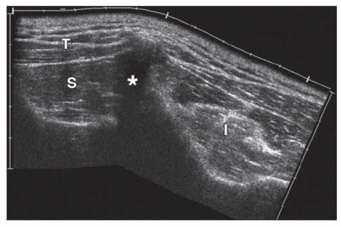
(S: supraspinatus muscles, T: trapezius muscles, I: infraspinatus, *: acoustic shadowing fromscabular spine)
The above shown figures are image result of EFOV; with a clear focus, the image shows clearly especially focusing on figure 1. We can be able to wrap up from the observation of these images that, the use EFOV technology is essential especially for musculosketal examination and it is evidently reliable. In fact, from the well elaborated figure using EFOV the images of supraspinatus and infraspinatus gives an opportunity to have a vivid picture of the structure and we can also be able have a comparison of the adjacent muscles.
3-D Ultrasound
The second technological advancement is that of the 3-D Ultrasound. The 3-D represents the three basic parts of the Ultrasound which are; data acquisition area, data analysis area and the 3-dimensional volume display (3D Ultrasound Imaging Group 2009). The 2-dimension images are acquired through by taking the images diagonally along the body surface in order to have a clear image; when the 2-Dimensional scan have been acquired they are then put in a computer software which is used to figure 3D images. There are a number of 3D imaging which offers a wide variety of focus which may facilitate to detect cancerous and benign tumors, examining of sensitive cases such as detecting prostates gland for early discovery of tumors, assessing the colon and rectum, imaging a fetus to evaluate its growth especially to observe cases of abnormal growth and so on (Freudenrich 2008).
Example of clinical study of 3-D Ultrasound
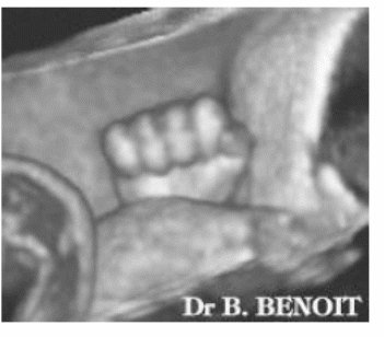
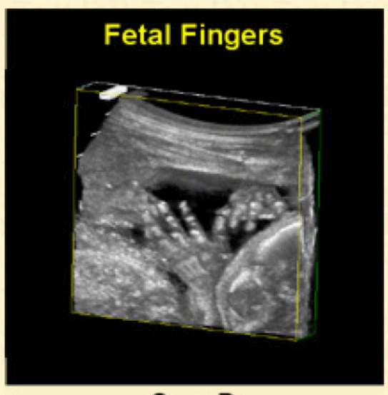
One of the basic applications of 3-D imaging is focusing development of foetuses. With the use of 3-D imaging, high and quality images of foetus can be acquired. In figure 5 provided in this paper, it is clearly observable that 3-D Ultrasound imaging has produced vivid images that can be used to draw a good contrast between the imaged tissues and the surroundings structures, such as, fluid and tissue.

Conclusion
Generally, there are numerous advancements in the real time imaging with two chief developments having taken place in the last decade. Extended field-of-View and 3-D Ultrasound technologies represents the two most significant developments and application these technologies in the society has seen a great revolution in the department of nursing. The two techniques can offer image with high resolution and display vivid images. EFOV can provide a huge display in one single image while the 3-D Ultrasound is widely helpful in focusing foetus. With the changing trends of technology, the future looks more promising and we might see more advancement of real time imaging for social change.
List of references
3D Ultrasound Imaging Group. 3D/4D Ultra. Web.
Antony, J. Ultrasound image gallery, A free gallery of high-resolution, ultrasound, color doppler and 3D images. 2007. Web.
Freudenrich, C. How Ultrasound Works. 2008. Web.
Kavanagh, E, and G koulourous. “Does extende field-of-view sonography improve interrater relaiability for the detection of rotator cuff muscle atrophy?” American Roentgen RAY Society 190 (2008): 27-31.
Kim, S, B Choi, K Kim, K Lee, and J Han. “Extended field-of-view sonography,advantages in abdominal applications.” Ultrasound Med 22 (2003): 385-394.
Nicholas, B. The Development of Ultrasound Machines. 2010. Web.
Poon, T, and R Rohling. “Three-dimensional extended filed of view Ultrasound.” World Federation For Ultrasound in Medicine and Biology (Elesevier) 32, no. 3 (2005): 357-369.
Srbely, J. “The Biophysical Effects of Ultrasound,a review of the current literature.” 2009, 3.