Introduction
Advancement of technology especially in the field of medicine has led to the invention of the nanobiosensor, which is a very powerful devise and a breakdown of a biosensor that comes in handy in recognising any “biochemical and biophysical signal associated with a specific disease at the level of a single molecule or cell” (Chen 2004, p.512). Nanobiosensor is therefore a mode of sensor that uses the nano skill to detect the presence of certain biological elements using chemical emissions that can be picked by the biosensor (Wilkins 1999: 600). In simple terms, it converts biological responses into electrical signals and hence a departure of the traditional methods that have been used over a long period using chemical reagents in laboratories (Rozlosnik 2009: 640). Thus, the technology can be used in the medical field, industries, and the military, as well as in domestic matters. Nanobiosensors can be used in the medical world to do the following things:
- It can be used in diagnostics in such a way that they detect the presence of certain diseases without the need for medical practitioners to take such samples like blood in some cases like diabetes tests
- It can be used in the manufacture of drugs whereby nanotechnology is used to separate different specific elements that are microscopic and or needed to produce a certain drug.
- Nanobiosensors can be used in industries especially in the food and drink industry. This technology is used in the process monitoring as well as control (Shipway 2000: 994: Ann 2008: 1010)). In military and environmental monitoring, the technology is used in the detection of poisonous gases and other elements.
The Components of (nano) Biosensors and their Functions
Or
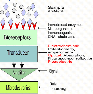
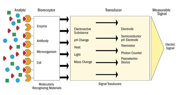
As evidenced by the two diagrams, the nanobiosensor is made of three distinct components in terms of functionality: the bioreceptor, transducer, and the detector.
Bioreceptor
A bioreceptor is a biomolecule that identifies the analyte or the chemical component that is being sought after in the test. Bioreceptors are molecularly recognised materials that have certain chemical elements that are used to find out the presence of specific elements. Examples of different bioreceptors can be enzymes, antibodies, and microorganisms. The bioreceptor’s work is to “bind the analyte to the sensor for measurement” (Gao 2007: 74). In addition, the bioreceptor has some elements that can be deduced by the transducer to come up with a signal that is indicative of the presence of the element being sought.
Transducer
The transducer is responsible for converting the chemical element from the bioreceptor and interpreting it into readable signals. The transducer is a form of interface that picks chemical change reactions by the bioreceptor for conversion into some energy that can be transmitted electrically to a detector that will give a form of result that can be interpreted to mean something (Luxton 2001: 990). When a component that is being measured is placed on the bioreceptor, it will cause a reaction in the atoms of the component thus producing energy signals that are converted by the transducer and then transferred to the detector, which is fashioned in a way that will give out an indicative signal.
Detector
The function of the detector is to give out data that can be interpreted to mean either a positive or a negative outcome. After the transducer has converted the chemical energy into electric energy, detectors’ function is to give out a signal that, when viewed by a person trained to use it, the person will be able to tell the results of the test. Detectors show results in the form of lights or digital numbers that will be indicative of the availability of a substance or the absence of the substance depending on the settings of the detector (Aroca 2004: 10274). Some detector might show the availability of the substance though failing to indicate whether the substance being tested is intense or just mild.
An overview of the detection principles: how each principle works
Nanobiosensors are mainly differentiated by the different bioreceptors that make them. The difference in bioreceptors is usually found in the molecular materials that it is meant to recognise.
Piezzo electric nanobiosensors
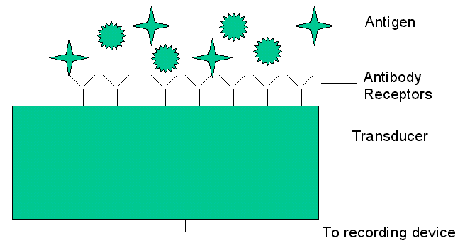
There are different detection principles available. Firstly is the principle of piezzo electric biosensors that measure changes in mass use elements such as quartz, which will vibrate when exposed to an electric filed. Other piezzo electric biosensors use gold as the detector to capture the angle at which electron waves are emitted when the element is exposed to laser light. The piezzo electric biosensor uses the principle that states, “Change in frequency is proportional to the mass of absorbed material” (Kundu 2010: 1113). Secondly, the principle of electrochemical biosensors applies when it comes to measuring changes in electrical distribution. It is believed to be the most receptive diagnostic gadget in place so far because of its ability to sense antigens in picogram scope. It can also sense “antigens in the gas phase as well as in the liquid phase” (Kundu 2010: 1113). An example of this is the immunosensor device.
Electrochemical Nanobiosensors
As Herna´ndez-Santos et al. Point, “Electrochemical nanobiosensors have mainly been fabricated from metallic nanoparticles” (2002: 1228). According to recent studies, “Metal nanoparticles have been used to catalyse biochemical reactions thus coming in handy in biosensor design” (Herna´ndez-Santos et al. 2002: 1229).
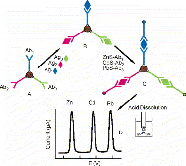
From the diagram above, (A) is “Introduction of antibody-modified magnetic beads, (B) is the binding of the antigens to the antibodies on the magnetic beads, (C) is the capture of the nanocrystal-labeled secondary antibodies, and (D) is the dissolution of nanocrystals and electrochemical stripping detection” (Chemla et al. 2000: 14270). Most of the studies done on the use of metal nanoparticles are mainly in catalysis. According to Curri et al., nano sized semiconductor crystals can also increase efficiency of photochemical reactions. They can be effectively coupled to biomolecular units such as enzyme to generate novel photo electrochemical systems (2002: 451).
- Magnetic nanobiosensors
Magnetic nanoparticles have found their use mainly in medical diagnostics and biology where they are used as powerful diagnostic tools (Chemla et al. 2000: 14270). According to Chemla et al., these nanoparticles may be prepared in different forms including “single domain or super paramagnetic (Fe3O4), greigite (Fe3S4), maghemite (g-Fe2O3), and various types of ferrites (MeO_Fe2O3, where Me = Ni, Co, Mg, Zn, Mn, etc.)” (2000: 14271). Cells are magnetically labelled with the magnetism being used to separate or isolate the cells. Another technique utilising magnetic nanoparticles is the magnetic immunoassay technique (Richardson et al. 2001: 992). A magnetometer is used to detect the targets that are magnetically labelled in the sampled to be assayed thus providing accurate information on qualities such as concentration. The use of magnetic nanobiosensors has been extensively studied with new techniques using sue paramagnetic nanoparticles and a magnifying device “based on a high-transition temperature dc Superconducting Quantum Interference Device (SQUID) (Chemla et al. 2000: 14270: Sarah 2004: 590). - Acoustic wave nanobiosensors
Acoustic wave nanobiosensors are some of the most sensitive nanosensors that demonstrate a marked improvement in the limits of detection (Sotiropoulou et al. 1996: 213). One variant of this technology is the mass-amplified quartz crystal microbalance assay that is utilised in a number of biological tests.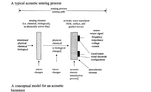
Source: (Sotiropoulou et al. 1996: 213) - Optical Nanobiosensors
Optical nanobiosensors come in two different forms. The colorimetric type detects changes in light absorptions. On the other hand, there is the photometric biosensor, which uses light intensity to produce an outcome that can bring out a certain meaning when interpreted (Tuan 2005: 919).An example of an optical nanobiosensor is the porous silicon optical biosensor (Psi), which is used to carry out whole blood tests. It works by size excluding cells and proteins larger than pore against getting into contact with the transducer surface
Conclusion
This technology can be useful when gadgets using nanotechnology are produced in masses for use in hospitals especially in developing countries where disease control is difficult. Nano science involves being able to see and sometimes control sheer minute particles that come in the form of atoms and molecules. Nano as a measure of the scale is measured to the scale of 1billionth of a meter denoted as 1nm (Tuan 2007: 411). Nano is measured using the nano scale. This can be crucial in the detection of dangerous nuclear particles on the earth surface that might be in minute form. Since nanobiosensors employ the nano expertise by using the atomic scale that counts in nanometres (Chen 2004: 508: Foultier 2005: 8), they have different applications. Their function is primarily to detect elements in a substance based on their atomic and molecular composition as well as distribution. This detection can be used in environmental monitoring for different elements that cause pollution. Nano technology and nanobiosensor can be of great help to the world of science due to its varied application for different purposes for the testing of different elements.
References
Ann, O, ‘Surface immobilisation methods for aptamer diagnostic applications’, Analytical and Bioanalytical chemistry, vol. 390, no. 4, 2008, pp. 1009-1021.
Aroca, F, ‘Gold nano particle embedded, self sustained chitosam films as substrates’, Langmuir, vol. 20, 2004, pp. 10273-10277.
Chemla, R, et al., ‘Ultrasensitive magnetic biosensors for homogeneous immunoassay’, Proc Natl Acad Sci U S A, vol. 97 no. 4, pp.14268– 72.
Chen, J, ‘Nano technology and biosensors’, Biotechnology advances, vol. 22, 2004, pp. 505-518.
Curri, L, et al. ‘Development of a novel enzyme/ semiconductor nanoparticles system for biosensor application’, Mater SciEng, C, Biomim Mater, SensSyst, vol. 22 no. 2, 2002, pp. 449– 52.
Foultier, B, ‘Comparison of DNA detection methods using nano particles and silver enhancement’, Nanobiotechnology, vol. 152, no. 1, 2005, pp. 3-12.
Gao, D, ‘Detection of tumour markers based on extinction spectra of visible light passing through gold nanoholes’, Applied Physics, vol. 90, no. 7, 2007, pp. 73-91.
Herna´ndez-Santos, D, Gonza´lez-Garcı´a, B, Garcı´a, C, ‘Review: metal-nanoparticles based electroanalysis’, Electroanalysis, vol. 14 no. 1, 2002, pp. 1225–35.
Kundu, T, ‘Development of evanescent wave absorbance-fibre optic biosensor’, Journal of Physics, vol. 75, no. 6, 2010, pp. 1099-1113.
Luxton, R, ‘The use of coated paramagnetic particles as a physical label in a magneto immunoassay’, Biosens bio electron, vol. 16, 2001, pp. 989-993.
Richardson, J, Hawkins, P, & Luxton, R, ‘The use of coated paramagnetic particles as a physicallabel in a magnetoimmunoassay’, Biosens Bioelectron, vol. 16 no. 1, 2001, pp. 989– 93.
Rozlosnik, N, ‘New directions in medical biosensors employing Polyderivative based electrodes’, Analytical and Bioanalytical chemistry, vol. 395, no. 3, 2009, pp. 637-645.
Sarah, M, ‘Biosensors for environmental monitoring’, Analytical and bioanalytical chemistry, vol. 378, 2004, pp. 588-598.
Shipway, A, ‘Nanostructured gold colloids electrodes’, Advanced materials, vol. 12, no. 13, 2000, pp. 993-998.
Sotiropoulou, S, Gavalas, V, Vamvakaki, V, Chaniotakis, A, ‘Novel carbon materials in biosensor systems’, Biosens Bioelectron, vol. 18 no. 5, 2003, pp. 211– 5.
Tuan, V, ‘Biosensors and biochips’, Biomemems and Biomedical nanotechnology, vol. 4 no. 1, 2007, pp. 410-431.
Tuan, V, ‘Fibre optic nanosensors for single cell monitoring ‘, Analytical and Bioanalytical chemistry, vol. 382, no. 4, 2005, pp. 918-925.
Wilkins, E, ‘Biosensors for detection of pathogenic bacteria’, Biosensors and bioelectronics, vol. 14, 1999, pp. 599-624.