Introduction
The cranial nerves are a group of 12 pairs of nerves located in the back of the human brain. They transmit electrical information between a person’s brain, face, neck, and torso. Cranial nerves aid in the senses of taste, smell, hearing, and touch (Smith & Border, 2019). If information is conveyed from the brain to the exterior, the nerve is efferent (motor). If the neuron passes from the extremities to the brain, it is an afferent (sensory) nerve. Sensory neurons are the 12 peripheral nervous system fibers that arise from the cranium’s foramina and fissures. Their number order (1-12) is dictated by the placement of their skull departure (rostral to caudal) since all of them start in the brain’s nucleus.
Discussion
Two are located in the frontal cortex (Olfactory and Optic), one has a component in the spinal column (Accessory), and the rest are located in the brainstem. Cranial nerves provide sensory and motor impulses to the tissues of the head and neck, hence regulating their functioning. Only the vagus nerve penetrates beyond the neck to perfuse the viscera of the thorax and abdomen. Table 1 below shows the twelve cranial nerves in their order number.
Table 1: List of the Twelve Cranial Nerves
Olfactory Nerve (Cranial Nerve One)
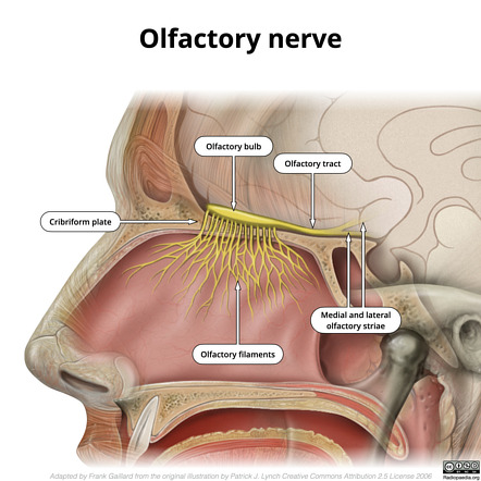
The numerous extensions of the olfactory nerve, known as fila olfactoria, travel via the cribriform plate of the ethmoid bone from the nasal cavity (Crespo et al., 2019). They conclude in the olfactory bulb, from which the olfactory tract proceeds. The olfactory tract fibers spread and terminate in the brain’s olfactory cortex (Crespo et al., 2019). Its neurons are located in the olfactory region, the nasal mucosa that borders the ceiling of the nasopharynx.
Optic Nerve (Cranial Nerve Two)
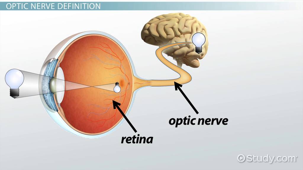
Everyone’s optic nerves travel through the bone-lined optical tube from the matching retina to the brain. The right optic nerve is derived from the right eye, while the left optic nerve is derived from the left eye. Optic fibers within the brain converge at the occipital lobe, a region just behind the pituitary gland (Smith & Czyz, 2021). Behind the skull, the nerves separate and send information to the right and left occipital hemispheres.
Oculomotor Nerve (Cranial Nerve Three)
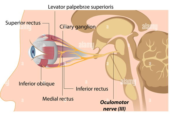
Each oculomotor nerve originates in the center of the brain, which is the upper portion of the cerebellum. A person’s oculomotor nerve passes to the eye on the exact hemisphere as the neuron through the cavernous sinus, a bone tube (Raza et al., 2018). The oculomotor nerve differentiates into numerous branches, each transmitting signals to a specific muscle.
Trochlear Nerve (Cranial Nerve Four)
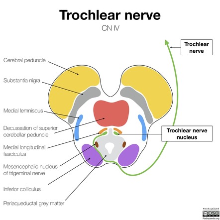
The trochlear nerve of an individual originates from the midbrain, underneath the position of the oculomotor nerve. This fiber powers the superior oblique tendon by traveling to the individual’s ipsilateral (same-side) eyeball (Agarwal et al., 2020).
Trigeminal Nerve (Cranial Nerve Five)
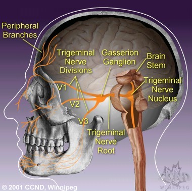
There are three somatic sensory extensions of the trigeminal nerve in humans: the ophthalmic nerve, the mandibular nerve, and the maxillary nerve. The ophthalmic nerve recognizes feeling from the upper region of the face, the maxillary nerve identifies sensibility from the central section of the face, and the mandibular division collects feeling from the bottom part of the face and has movement patterns (White et al., 2021). Underneath the midbrain, the trigeminal nerve exits from the pons Varolii to the hypothalamus.
Abducens Nerve (Cranial Nerve Six)
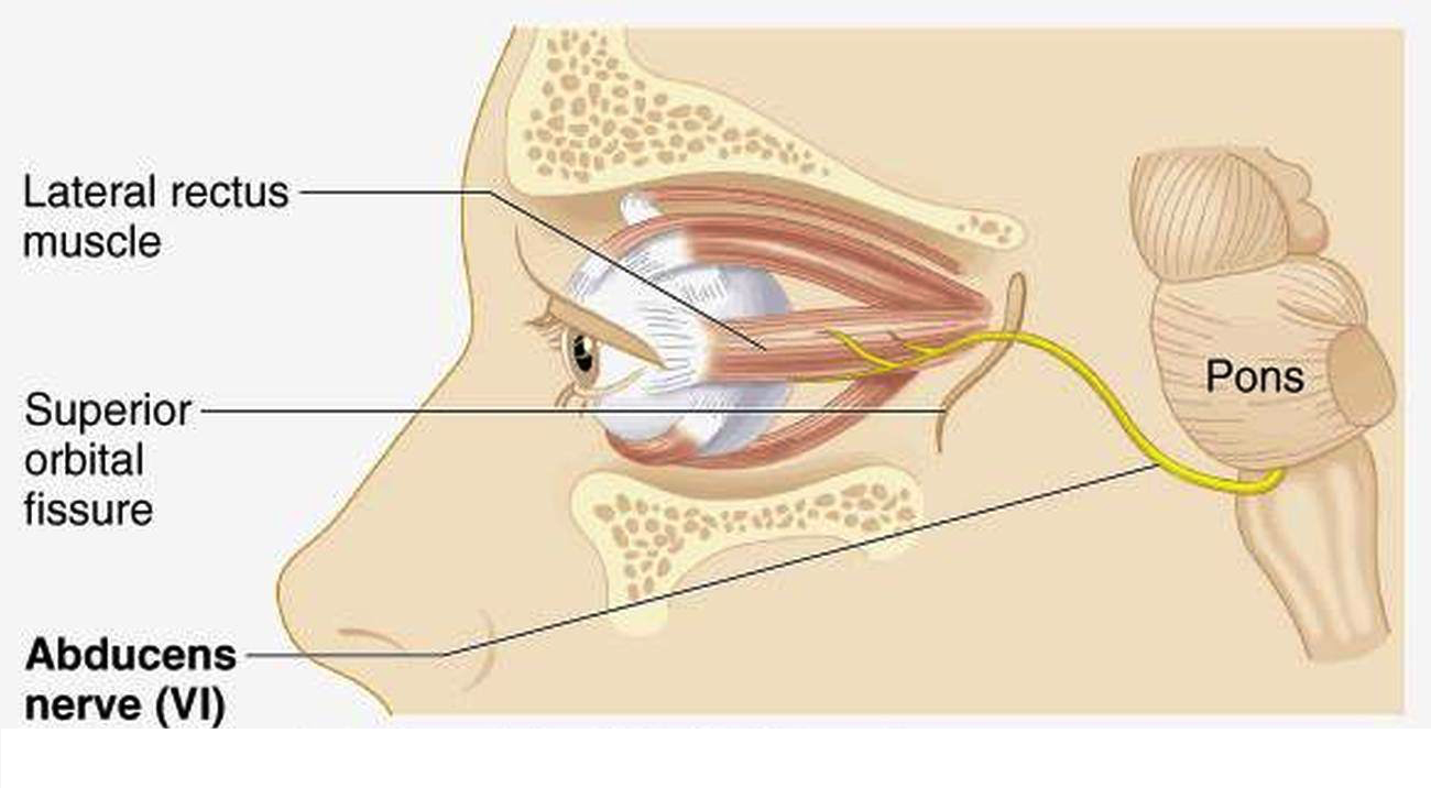
The lateral rectus tendon is innervated by cranial nerve 6, which is a broad somatic efferent neuron in the human body. This nerve arises from the inferior pons and extends toward the rectus abdominis muscle in the eye (Lucio et al., 2022).
Facial Nerve (Cranial Nerve Seven)
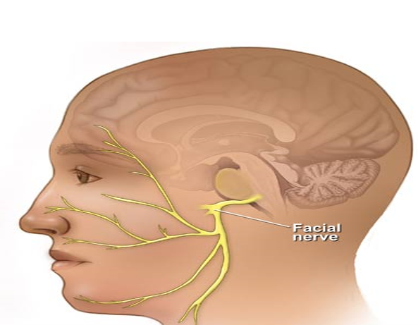
The seventh cranial nerve is a multifunctional nerve with general and specific components. A broader main stem containing motor neurons and a narrower transitional nerve containing perception and parasympathetic axons emerge from the cerebellum (Takezawa et al., 2018). The two segments exit the brain structure via the internal acoustic meatus and go through the facial tract. Together, they exit the skull via the stylomastoid foramen after joining to create the facial nerve proper (Takezawa et al., 2018).
Vestibulocochlear Nerve (Cranial Nerve Eight)
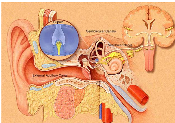
Vestibulocochlear nerve sensation neurons are situated in the inner ear and penetrate the lower portion of the pons together. The vestibular and auditory elements of the vestibulocochlear nerve obtain information depending on hair cells’ motion in the inner ear (Walijee et al., 2021). This data is employed to notify a person’s body of their position, allowing them to retain their equilibrium, transmit sound impulses to their brain, and interpret the noises they perceive.
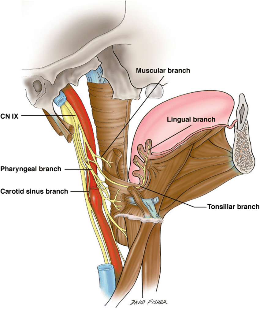
Glossopharyngeal Nerve (Cranial Nerve Nine)
The glossopharyngeal nerve originates from the hypothalamus, the lowest portion of the cerebellum positioned above the spinal column, and descends to the throat and mouth (García Santos et al., 2018).
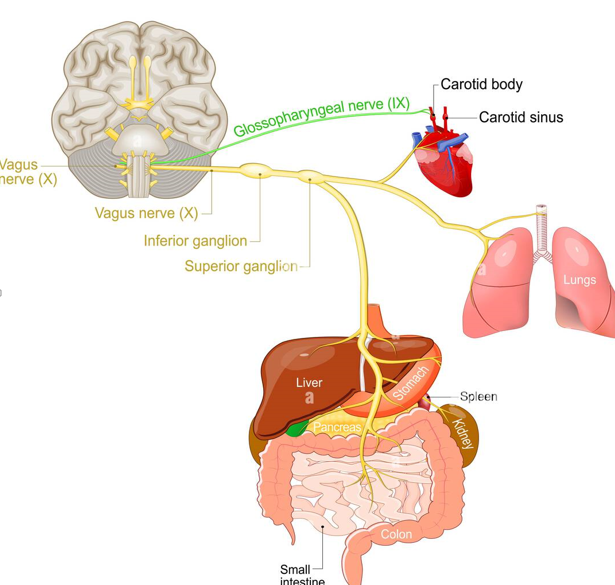
Vagus Nerve (Cranial Nerve 10)
The vagus nerve arises from the medulla and proceeds beside the carotid artery in the neck, outside the skull. In addition, the vagus nerve separates into segments that travel to the heart, digestive tract, and lungs (Butt et al., 2020).
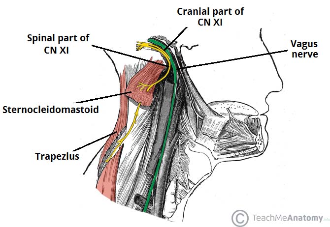
The auxiliary nerve assists in raising the shoulders and rotating the head and neck. This fiber emerges from the brainstem and descends toward the sternocleidomastoid and trapezius musculature outside the cranium (Johal et al., 2019).
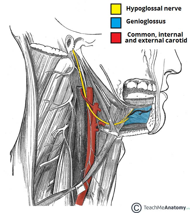
Hypoglossal Nerve (Cranial Nerve 12)
This nerve coordinates the mobility of a person’s tongue with their capacity to speak and gulp down and is located in the base of the tongue. Following its beginning in the cerebellum, the hypoglossal nerve descends beyond the oral mucosa to reach the skeletal muscles of the tongue (Iaconetta et al., 2018).
References
Agarwal, N., Ahmed, A. K., Wiggins III, R. H., McCulley, T. J., Kontzialis, M., Macedo, L. L., Choudhri, A. F., Ditta, L. C., Ishii, M., Gallia, G. L., Aygun, N., & Blitz, A. M. (2021). Segmental imaging of the trochlear nerve: Anatomic and pathologic considerations. Journal of Neuro-Ophthalmology, 41(1), 7-15.
Butt, M. F., Albusoda, A., Farmer, A. D., & Aziz, Q. (2020). The anatomical basis for transcutaneous auricular vagus nerve stimulation. Journal of anatomy, 236(4), 588-611.
Crespo, C., Liberia, T., Blasco‐Ibáñez, J. M., Nácher, J., & Varea, E. (2019). Cranial pair I: The olfactory nerve. The Anatomical Record, 302(3), 405-427.
García Santos, J. M., Sánchez Jiménez, S., Tovar Pérez, M., Moreno Cascales, M., Lailhacar Marty, J., & Fernández-Villacañas Marín, M. A. (2018). Tracking the glossopharyngeal nerve pathway through anatomical references in cross-sectional imaging techniques: A pictorial review. Insights into Imaging, 9(4), 559-569.
Iaconetta, G., Solari, D., Villa, A., Castaldo, C., Gerardi, R. M., Califano, G., Stefania, M., & Cappabianca, P. (2018). The hypoglossal nerve: anatomical study of its entire course. World Neurosurgery, 109, 486-492.
Johal, J., Iwanaga, J., Tubbs, K., Loukas, M., Oskouian, R. J., & Tubbs, R. S. (2019). The accessory nerve: A comprehensive review of its anatomy, development, variations, landmarks and clinical considerations. The Anatomical Record, 302(4), 620-629.
Lucio, L. L., Freddi, T. D. A. L., & Ottaiano, A. C. (2022). The Abducens nerve: Anatomy and Pathology. In Seminars in Ultrasound, CT and MRI. WB Saunders.
Raza, H. K., Chen, H., Chansysouphanthong, T., & Cui, G. (2018). The aetiologies of the unilateral oculomotor nerve palsy: A review of the literature. Somatosensory & Motor Research, 35(4), 229-239.
Romano, N., Federici, M., & Castaldi, A. (2019). Imaging of cranial nerves: A pictorial overview. Insights into imaging, 10(1), 1-21.
Smith, A. M., & Czyz, C. N. (2021). Neuroanatomy, cranial nerve 2 (Optic). In StatPearls. StatPearls Publishing.
Smith, C. F., & Border, S. (2019). The twelve cranial nerves of christmas: Mnemonics, rhyme, and anatomy–seeing the lighter side. Anatomical Sciences Education, 12(6), 673-677.
Takezawa, K., Townsend, G., & Ghabriel, M. (2018). The facial nerve: Anatomy and associated disorders for oral health professionals. Odontology, 106(2), 103-116.
Walijee, H., Vaughan, C., Munir, N., Youssef, A., & Attlmayr, B. (2021). Microvascular compression of the vestibulocochlear nerve. European Archives of Oto-Rhino-Laryngology, 278(10), 3625-3631.
White, T. G., Powell, K., Shah, K. A., Woo, H. H., Narayan, R. K., & Li, C. (2021). Trigeminal nerve control of cerebral blood flow: a brief review. Frontiers in Neuroscience, 15, 1-9.