Cardiovascular technology involves the use of mathematic modeling which has been a part of physics from the days of Isaac Newton (Van de Vosse, 2003, p. 175). Though the fact is accepted, the physical difficulty in acquiring biological systems for experimentation allows mathematic modeling to be used only sparingly in human technologies. Medical treatment is also more dependent on behavior of the body rather than predictive modeling. However modern times have witnessed the progress of mathematic to computational and analytic mathematics. Due to the simultaneous progress in understanding the human biological systems and innovative imaging techniques, mathematic modeling has become an integral part of modern health care and could predict the internal and external changes in the human body.
Cardiovascular Technological Innovations
The artificial heart is a dream on which various cardiac researchers have worked upon. The auxiliary pneumatically-driven pulsatile assist pump has been installed in place of the heart in about 700 patients in Japan (Umezu, 2007, p.88). Then a clinically better quality ventricular assist pump, called EVAHEART was made and implanted in 11 cases, the first one in 2005. The first 3 patients have been alive for 2 years now. Bioengineers have been employing the basics of phsyics and calculations for in vitro hydrodynamic performance tests apart from other tests to come up with the artificial hearts. 9L/mt of blood pump flow was achieved with EVAHEART when the blood pressure was 100mm.Hg. and the pump speed was 2400rpm which compared to a satisfactory bypass flow.
Continuing developments in cardiovascular technology as in any other medical field and the emergence of newer imaging technologies have revolutionized cardiac diagnosis and therapy.
“Ultrasound examination including echocardiography, scintigraphy using single photon and positron emitting radiopharmaceuticals, magnetic resonance with or without a paramagnetic imaging agent, and by X-ray computed tomography or cardiac catheterisation and angiography with the injection of an iodinated contrast agent” have all been used (Fraser et al, 2006, p. 955). Diagnostic cardiac catheterization is also being done on a regular scale. Percutaneous intervention makes coronary arteriography a fairly easy procedure which also incorporates treatment. Abnormal haemodynamic function needs be ascertained only at the time of procedure. and immediate intervention is made if necessary. The usefulness of physics is obvious here.
Coronary vessel visualisation is possible by using a 16 row Multi-slice computed tomography (MSCT) scanners with retrospective ECG-gating (Kuettner, 2005, p. 331). Cardiovascular MRI allows the visualisation of not only the anatomy of the heart but also its function, metabolism, perfusion, and, more recently, proximal coronary artery status (Feliu, 2008, p. 27). Cardiac magnetic resonance (CMR) is emerging as a technique for studying congenital and acquired cardiac pathology. Multidetector CT (MDCT) coronary angiography is another indispensable tool. Myocardial ischaemia and injury is examined by the single photon emission computed tomography, cardiac magnetic resonance or the contrast enhanced MRI (Feliu, 2008, p. 29).
Biomechanics
Fung, one of the founders of modern biomechanics, defined it as the mechanics applied to biology: the research discipline that studies the mechanical properties of organisms and helps in understanding of their normal and pathological function, helps to predict their adaptation to changing circumstances and helps in finding methods for artificial intervention (1993).
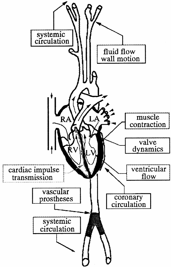
Cardiovascular biomechanics focuses on the heart and the blood vessels of the cardiovascular system (CVS). The activities of the CVS include the pumping of the heart muscle and valves, the dynamics of blood flow, the wall motion of the blood vessels and heart and the exchange between the nutrients and oxygen with the excretory products and carbon dioxide in the various organs. Cardiovascular biomechanics is significant in the diagnostics, therapy selection, surgery and other interventions. This research area includes solid and fluid mechanics of structures complex in nature and essential to the human life (Van de Vosse, 2003, p. 176).
The figure on the next page shows a schematic representation of the CVS (Van de Vosse, 2003, p.177). Mechanically speaking, the heart is a four chambered pump which causes the circulation of blood through the body.
The heart muscle like any other muscle has depolarization waves which transmit the cardiac impulses and cause contraction. The two valves between the atria and ventricles on the left and right sides allow unidirectional blood flow like the main arteries connected to the heart, the pulmonary and aorta. The systemic circulation taking oxygenated arterial blood to the different organs, the capillary system in these tissues where exchange of the oxygen and nutrition products with the excretory products occurs and the venous blood returning blood to the heart for cleansing all toxic products from the circulatory system. This physiological functioning is regular and highly optimized (Van de Vosse, 2003, p. 176).
Mortality from heart disease is more than 40%. The illnesses of the CVS include atherosclerosis causing progressive narrowing of vessels at particular spots due to plaques which lead to partial or total occlusion and can lead to embolism and thrombosis or ischaemia which could be fatal (Giddens et al, 1993). Heart failure due to disturbed contraction patterns, stenosis or leakage of the valves or hypertension could occur.

Diagnostic methods include the X-ray, the CT (Computed Tomography), the US (ultrasonography) and the MRI (Magnetic resonance imaging) provide geometric information and even quantitative measures of flow. Wall strain or stress cannot be directly measured but can be predicted by mathematical models. The diagnostic measurements and the mathematical modeling together constitute simulation-based diagnostics (Van de Vosse, 2003, p. 177).
Atherosclerosis is often treated with partial replacement, reconstruction, bypassing with prosthesis or autografts, balloon angioplasty with or without stents. The procedures have saved many lives but they have yet to reach foolproof standards and may have to be repeated. Better biomechanical knowledge would change matters. Mechanical functioning and biocompatibility
of mechanical, biological and tissue engineered prostheses have to be further improved. Mathematical modeling also has provided for methods to monitor the function and adaptation of the heart muscle after therapy or interventions like the effect of valve replacement, application of cardiac-assist devices or pace makers.
The mathematical models should be able to provide information about the physiological and pathological phenomena that occur and also give patient-specific information so that the clinical diagnosis improves and the patient may be provided with a good therapeutic intervention. The latter is possible through medical imaging and function measurements (Van de Vosse, 2003, p. 178).
The pressure and flow waves in the pulmonary and systemic arterial systems are caused by the contraction of the heart muscle based on the pressure and volume of the blood in the chambers and these actions are dependent on the perfusion of the contractile heart muscle by the coronary circulation. To rule out the possibility of a flaw in the dynamics by the closing and opening of the valves and inertia forces because of flow in the left ventricle, the contraction mechanism has an added electrical cardiac impulse transmission. The whole set of systems is again influenced by the central, local regulatory and adaptive mechanisms of the heart and arterial wall ( Milnor, 1990). The mathematical models that have been suggested by various authors do not embody the total concept. The researches only provide piecemeal information.
Two examples of mathematic models for the CVS (Broz, Mathematic modeling of the cardiovascular system).
Chemical and Mechanical performance
Changes in the concentration of calcium ions regulates the contractility of the heart muscle. This produces pressure pulsations. The Beeler-Reuter equations describe the membrane potential that affect the concentration of calcium ions transported through the cardiac muscle membrane. The potentials in the action of the atria and ventricles depend on four ionic currents.
- INa – fast inward sodium current
- IS – slow inward calcium current
- IK – time independent potassium current
- IX – time dependent outward potassium current

where Cm[μF/cm2] is the capacitance of myocardial tissue and Ist(i)[μA/cm2] is the stimulating
current. The ionic currents are formulated in this way :


The pressure in the heart is described by the equation derived from the general energy and entropy balance :

where the intracellular concentration of calcium ions cCa is calculated from the following differential equation :
Hemodynamic performance
Following equation describes pressure changes in the pulmonary artery and aorta

i=2,8 (8).
For other segments in pulmonary and systemic circuit we can omit the last item in the equation and use it in following form :
i=3,4,5,9,10,11,12,13,14,15 (9).
where C[m3/Pa] denotes the compliance, VU[m3] is the residual volume and V[1] represents the wall viscosity. Pressure in dialysis pump is described by following equation :
Blood flow between compartments is determined by the balance of momentum in the following differential and ordinary equations:
Between ventricles and output arteries, and between dialysis pump and systemic veins:

j=1,7,16; k=2,8,11 (11)
In flow that comes from pulmonary artery or aorta we can omit the last item that represents blood inertia:
j=2,8,8;
k=3,9,12 (12).
flow between other segments are described in following form:

j=3,4,9,9,9,9,10,13,14,15;
k=4,5,10,14,15,16,11,11,11,11 (13).
or
j=0,5,6,11,12 ;
k=1,6,7,0,13 (14).
where L[Pa.s2/m3] characterizes the blood inertia, R[Pa.s/m3] is the hydrodynamical resistance, [1] is the coefficient of blood inertia and A[m2] is the flow area. The volume changes in all segments of the cardiovascular system are determined by the balance of mass.
i, j, k=0,1,2,…,16 (15)
Conclusion
Knowledge of biomechanical aspects is important for understanding the patho-physiology of the CVS. The development and interpretation of diagnostic data also is possible.
Mathematic modeling has in addition a significant role in CVS diagnostics, the development and optimization of prostheses and medical devices, prediction of outcome of intervention and in the selection of therapy and intervention planning. Mathematic modeling is yet to reach a wholesome model. Deliberate decoupling of the functions of the CVS may provide models which are good for some functions only. Cardiovascular technology is very dependent on mathematical modeling and thereby physics for its future innovative technologies.
References
Broz, 2009, Mathematic modeling of the Cardiovascular system. Web.
Feliu, E. et al. (2008). “Cardiovascular Imaging Chapter 2 in (Eds ) Learning diagnostic Imaging by R.Ribes, Luna A. and Ros. P.R., Springer Verlag, Berlin Heidelberg.
Fraser et al, (2006). “The future of cardiovascular imaging and non-invasive diagnosis” Eur J Nucl Med Mol Imaging 33:955–959. Web.
Fung, Y.C. (1993) “Biomechanics: Mechanical Properties of Living Tissues”. New York: Springer-Verlag (1993) 433 pp.
Giddens, D.P., C.K. Zarins and S. Glagov, (1993). “The role of fluid mechanics in the localization and detection of atherosclerosis. ASME J. Biomech. Eng. 115 (1993) 588–594.
Kuettner, A. et al, (2005). “Coronary vessel visualization using true 16-row multi-slice computed tomography technology”. The International Journal of Cardiovascular Imaging (2005) 21: 331–337.
Milnor, W.R. (1990). “Hemodynamics”. New York: Oxford University Press 501 pp.
Umezu, M. (2007). “How to Ensure Safety Factors in the Development of Artificial Heart: Verified by the Usage of “Modeling and Simulation” Technology in (Eds.) K.Li, Life System Modeling and Simulation 2007, LNBI 4689, pp. 88–96. Springer- Verlag Berlin Heidelberg.
Van De Vosse, F.N. (2003). “Mathematical modelling of the cardiovascular system”. Journal of Engineering Mathematics 47: 175–183. Kluwer Academic Publishers. Printed in the Netherlands.