Updated:

Chemical Evidence for a Prokaryotic Origin of Subcellular Organelles Essay

Exclusively available on Available only on IvyPanda® Made by Human No AI

Introduction

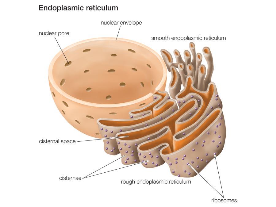
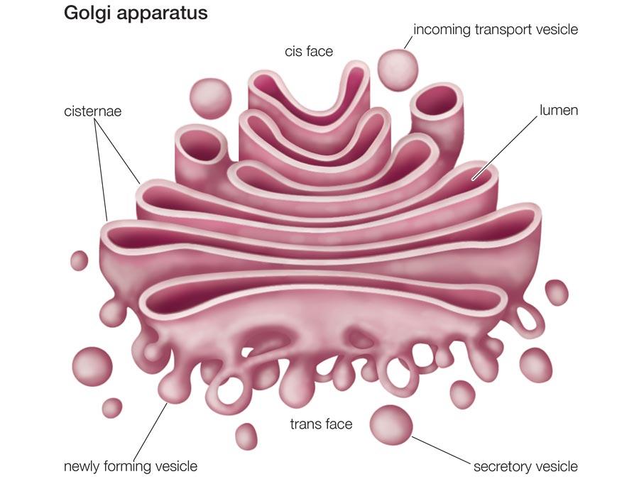
An organelle is a structure within a cell specialized to perform specific activity or function. An organelle is also enclosed by a specialized membrane known as lipid bi-layer. There are many organelle types within eukaryotic and prokaryotic cells. Major organelles include the nucleus, mitochondrion, ribosomes as well as chloroplasts. Other significant organelles include the Golgi apparatus, endoplasmic reticulum, lysosomes in addition to vacuoles.

Compared to prokaryotes, eukaryotes have many and more complex organelles. The word organelle is derived from the word organ. This is because the organelle has a specific activity in the cell as an organ has a specific function in the body. In the early 1920’s scientists used the term organelle while describing propulsion structures; examples of such includes the flagella in addition to their anchors. Another example includes protists structures such as ciliates.

As science advanced, the term organelle was used by scientists to describe cellular structures enclosed by a membrane that was in the 1950’s. Today organelles refer to structures within the cell that perform specialized activities and are enclosed by a lipid membrane. There are organelles responsible for energy production; main organelle involved includes the mitochondrion. Another example includes the chloroplasts whose function is production of chlorophyll which is vital in photosynthesis.

Prokaryotic organelles

Prokaryotes are less complex than eukaryotes metabolically and structurally. Early scientists believed that prokaryotes contained no membrane-enclosed structures. However; research has proved that prokaryotes indeed have organelles. At one time, Prokaryotes were time and again viewed as organelles that were less endowed in terms of their internal organization.

Nevertheless, research into the internal structure of prokaryotes has already yielded details regarding the internal structure of these organelles. During the early 1970s, an idea was developed to the effect that bacteria did contain mesosomes; some kind of membrane folds. However, further researcher revealed that these were in fact production artifacts of the various chemical agents that were then in use for the preparation of bacterial cells prior to further study on an electron microscope.

Nonetheless, recent research studies have divulged the fact that some prokaryotes do indeed contain microcompartments. An example of such microcompartmets is carboxysomes. The microcompartments found in Prokaryotes tend to be subcellular in nature, with their diameter size ranging between 100 and 200 nm. In addition, the subcellular compartments are usually enclosed by a proteinaceous shell.

Evidence supporting this argument is the existence of magnetosomes. Despite the absence of nucleus within a prokaryotic cell, prokaryotes have structures resembling a nucleus; they are referred to as planctomycetes. These are normally surrounded by membranes made up of lipids.

The endosymbiotic theory

This theory is involved with plastids and mitochondrion origin. These all constitutes part of the eukaryotic cells’ organelles. The endosymbiotic theory holds that the plastids and mitochondrion came into being as distinct entities of prokaryotic organisms. It is believed that organelles were originally free living prokaryotes. This argument is supported by the endosymbiotic theory. Postulates of this theory are:

  • Aerobic bacteria are ancestors of modern day eukaryotic mitochondria. Ancestral aerobic bacteria are related to Rickettsias.
  • Eukaryotic chloroplasts originated from cyanobacteria.

Evidence supporting this theory

Mitochondria as well as chloroplasts rise from existing chloroplasts plus mitochondria. They cannot rise within a cell void of them since nuclear genes code only for proteins making them. Another proof supporting this theory is resemblance of bacterial genome with that of chloroplasts and s well as mitochondrion. Both organelle’s DNA and bacterial DNA have circular genomes. Both genomes lack histones thus their similarity.

Chloroplasts plus mitochondria have protein synthesizing mechanisms which is similar to that found in bacteria. They have ribosomes responsible for protein synthesis through transcription, translation and protein modification processes. These organelle’s first amino acid during transcription is fMet which is always the case in bacteria thus the similarity with bacterial protein synthesis mechanism.

Proof of endosymbiotic theory is depicted by various antibiotics such as streptomycin. This antibiotic is acts by curbing protein synthesis within bacteria and other prokaryotes. When administered, the antibiotics also blocks mitochondrial plus chloroplast protein synthesis process but spares protein synthesis within cytoplasm of eukaryotes. This shows similarity between these organelles and bacteria thus prokaryotic origin of both mitochondria plus chloroplast.

Protein synthesis inhibitors within eukaryotic cells do not affect protein synthesis within bacterial cells, mitochondria as well as chloroplasts. For example, diphtheria toxins affect eukaryotic cells by curbing protein synthesis; the toxin has no effect on bacteria cells and the two organelles. This shows similarity between bacterial cells and mitochondria plus chloroplasts.

Another proof to this theory is shown through administration of rifampicin which is an antibiotic. On administration, the antibiotic inhibits RNA polymerase within the bacteria, chloroplast as well as mitochondria however; the drug has no effect on RNA polymerase in eukaryotic nucleus. This shows similarity between these organelles and bacteria thus their prokaryotic origin.

Chloroplast

Chloroplast is responsible for photosynthetic activities within the cell. Chloroplasts have disc shape with 4-6 nm diameters. The internal structure of chloroplast includes the sroma and grana. Sroma consists of a network of disks known as thylakaoids. Thykaloids are extensively interconnected. When stacked together, thykaloid forms the grana. Thykaloids contain chlorophyll which is important for photosynthetic processes.

Chlorophyll absorbs light energy vital for photosynthetic processes. After absorption of light energy by chlorophyll, it is converted to chemical energy also known as adenosine triphosphate (ATP). Conversion of light energy to chemical energy is a complex process taking place within the grana. Apart from supporting photosynthesis, the grana stores photosynthetic products. Example includes starch which is the storage form of glucose within plants.

The stroma consists of enzymes as well as proteins necessary for conversion of Carbon dioxide as well as water into glucose and oxygen respectively. This is a very important reaction as it provides energy in form of carbohydrates. It also regulates the oxygen-carbon dioxide concentration in the environment. Proteins involved in photosynthetic reactions include the carotenoids which are found within the stroma.

Apart from producing photosynthetic products, the chloroplasts are responsible for synthesis of products such as lipids, oils, proteins as well as scents however; these products are produced in minute concentrations.

Chloroplast
Fig.1. Chloroplast.

Origin of chloroplast

There are several theories depicting the origin of chloroplasts. An example includes endosymbiotic theory. According to this theory, chloroplasts existed as independent or free living prokaryotic organisms. As a proof to this argument, chloroplasts consist of DNA. DNA contained within chloroplast is responsible for synthesis of chloroplast proteins which includes carotenoids.

Endosymbiotic theory also argues that chloroplasts once existed as photosynthetic bacteria. As time elapsed, this bacterium was engulfed within a nonphotosynthetic cell. In engulfed state, the two parties lived symbiotically where each needed the other party for metabolic purposes. With time, photosynthetic bacteria lost the ability to live independently through evolution. Scientists argue that what remained of photosynthetic bacteria is present day chloroplast organelle.

Evolution led to loss of large amount of photosynthetic bacteria’s DNA to host cell’s nucleus the eventual inability to live independently. Loss of much DNA to the host cell gave the host cell the ability to control functions of photosynthetic bacteria that later became chloroplasts.

Another proof supporting this theory is chloroplast’s DNA translation as well as composition. Chloroplast DNA is translated in a similar fashion as that found within bacterial cells. Its composition is also similar to DNA found within the bacteria. Due to this resemblance, endosymbiotic theory is significant in prokaryotic chloroplast origin.

Synthesis of chloroplasts within a cell occurs following a genetic code contained in the organism DNA. This process is summarized into three major steps namely transcription, translation and expression. This process involves coordination of various RNA; that is transfer RNA as well as messenger RNA. Each has significance in production of proteins used in production of chloroplasts.

The first step in protein synthesis is transcription. This is typically the transfer of genetic code from DNA strand to messenger RNA. The genetic code copied in this case is relevant to synthesis chloroplasts. Transcription process begins with separation of double stranded DNA. This process is initiated by RNA polymerase which is an enzyme necessary for production of mRNA. One DNA strand serves as a template for synthesis of mRNA.

The resultant mRNA has two regions that is introns and exons. Exon consists of significant coding regions while introns contain redundant sequences with no significance in protein synthesis. The last step in transcription process is splicing process where exons are linked together after removal of introns.

The next major step in synthesis of proteins is translation. In this process, sequences carried by messenger RNA are converted to relevant bases depending on protein being made. Base sequences produced leads to production of amino acids which code for specific proteins. This process takes place within ribosomes. Transfer RNA is very important as it enhances growth of amino acid strands that translates to synthesis of amino acids.

The next major important step taking place in synthesis of proteins is expression where additional modification takes place. Examples of modification include 5” end capping. With presence of DNA within the chloroplast show some sort of independence due to ability to synthesis proteins from DNA sequences thus the endosymbiotic theory.

Chloroplast’s genome

Chloroplast found within Marchantia polymorpha which is a liverwort has 121,024 base pairs which are enclosed within a circle. Within this genome, there are various genes coding for 23S, 16S, 4.5S, and 5S subunits. These subunits belong to ribosomal RNA important during protein synthesis. Within chloroplast genome, there exist thirty seven genes which code for transfer RNA which important during translation step of protein synthesis.

Within the genome, there exist a total of four genes which code for RNA polymerase important in transcription process in protein synthesis process. Chloroplast’s genome also has a gene responsible for coding RUBISCO enzyme. RUBISCO stands for ribulose biphosphate carboxylase oxygenase. Objective of this enzyme is addition of carbon dioxide to ribulose biphosphate which triggers the Calvin cycle within the chloroplast. The chloroplast similarity with that of bacteria shows clearly the origin of chloroplast from prokaryotes via endosymbiotic hypothesis.

Mitochondrion

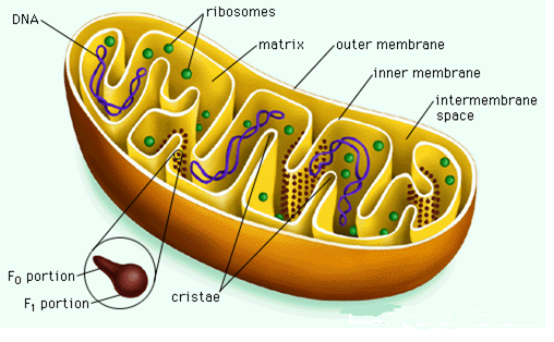
These are very important organelles within the cell because they are responsible for production of energy within the cells. This explains why cells requiring a lot of energy have many mitochondria within the cytoplasm example of such cells includes muscle cells in addition to cells containing flagella and cilia. It is within the mitochondria that ATP producing reactions takes place; example includes glycolysis as well as Kreb’s cycle. Mitochondria have a sausage shape as shown in the figure below.

Mitochondria 
Fig.2. Mitochondria.

Main features within the mitochondria are the bi-lipid membrane, cristae and s well as matrix. The bi-lipid membrane allows selective passage of elements and compounds. Cristae are the finger-like structures within the inner membrane layer. Cristae increase the surface area for energy production through aerobic respiration. The matrix consists of both enzymes as well as proteins necessary for aerobic respirations. Example of proteins found within the matrix includes cytochrome molecules.

Mitochondrial matrix

This is the engine of the mitochondria. It contains many components which vary from proteins to enzymes. Main activity taking place within the matrix is production of energy through citric acid cycle. Energy produced is in form of ATP through activation of an enzyme called ATP synthase. ATP synthase is located within the inner mitochondrial membrane.

Apart from proteins and enzymes, the matrix contains several copies of mitochondrial genome. Enzymes found in the matrix catalyze pyruvate as well as fatty acid oxidation processes.

Reactions within the matrix

Main reaction occurring within the matrix is production of ATP through the citric acid cycle. This cycle begins with transportation of pyruvate to matrix across mitochondrial membrane. Within the matrix, pyruvate is oxidized and mixed with coenzyme A. This results to production of acetyl-CoA, Carbon dioxide as well as NADH.

Production of Acetyl CoA is vital since it is the main substrate for citric acid cycle. Oxidation of acetyl CoA by enzymes within the matrix leads to formation of substrates used in electric transport chain. Example of these includes FADH2 as well as NADH. Other products produced include GTP that is converted easily to ATP. This mechanism is similar to that one depicted by aerobic bacteria thus a proof that mitochondria originated from prokaryotes.

Mitochondrial genome

Mitochondrial genome has a circular form similar to that of bacteria and other prokaryotes. The genome has a total of 16,569 base pairs coding for a number of genes. Examples of genes within the genome include genes coding for rRNA molecules, twenty two tRNA molecules as well as thirteen polypeptides. The thirteen polypeptides are responsible for building various proteins embedded within the inner membrane of mitochondria.

Other genes found in mitochondrial membrane code for cytochrome b, ATP synthase, cytochrome c oxidase as well as NADH dehydrogenase. These products are enzymes and proteins necessary for energy production within mitochondrial matrix. Mitochondrial genome resembles aerobic bacteria’s genome thus proving that mitochondrial originated from prokaryotes.

Additional mitochondrial functions

Apart from production of energy, mitochondria display a number of functions. These includes control of membrane potential, apoptosis which is also known as programmed cell elimination, control of cellular proliferation, control of cell metabolism, synthesis of steroids as well as synthesis of heme.

Origin of mitochondria

It is depicted that mitochondria originated from an ancestral aerobic bacteria. Mitochondrion poses DNA within its matrix which proves endosymbiotic theory. Presence of DNA within the matrix shows mitochondria existed as an independent organism before evolution occurred. Another proof showing prokaryotic origin of mitochondria is ability of mitochondria to divide through binary fusion just as prokaryotes such as bacteria.

Another evidence showing originality of mitochondria from prokaryotes is binary fission which is independent of host cell. This shows that mitochondria once existed as independent constituents before evolution took over. This argument is further explained by the figure below. It shows how symbiosis association led to evolution of aerobic bacteria to mitochondria.

Mitochondrial evolutions
Fig.3: Mitochondrial evolutions.

Due to these facts, many scientists support the endosymbiotic theory where prokaryotes with aerobic respiration capability were enclosed or engulfed by larger prokaryotes. This lead to symbiotic relationship between the two prokaryotes since the larger prokaryotes provided shelter and nutrients to the smaller prokaryotes while the smaller prokaryotes provided energy through aerobic respiration.

As time lapsed, the larger prokaryotes absorbed much DNA from the smaller prokaryotes thus controlling them both metabolically and functionally. Today, the smaller prokaryotes engulfed within large prokaryotes evolved to mitochondria. This explains its origin. Since the smaller prokaryotes had aerobic respiration capability, this explains mitochondria’s ability to produce energy through aerobic respiration. Energy in form of ATP is produced in the mitochondrion’s matrix which has enzymes necessary aerobic respiration.

Additional Evidence showing origin of mitochondria and chloroplasts from bacteria

There are numerous facts supporting rise of these organelles from prokaryotes. Mitochondria in addition to chloroplasts divide in a binary fission fashion which is similar to bacteria. The internal structure of both organelles resembles bacterial structure (Bruce et al, 2002).

Apart from structural similarity, the two depicts similarity in biochemical reactions. Ribosomes located within cyanobacteria resemble those found in both mitochondria plus chloroplasts. Their similarity lies with number of subunits. Plastids have a size that approximates the size of mitochondria.

Ribosomes

These are very important organelles as they are very relevant in synthesis of proteins. The name ribosome is derived from ribonucleic acid (RNA) because they are very rich in RNA. Ribosomes found within the prokaryotes differ considerably with ribosomes found within eukaryotes. For example, eukaryotic cells contain 4- RNA strands which are associated with seventy to eighty proteins.

On the other side, prokaryotes have ribosomes linked with 3 strands of RNA associated with fifty proteins. Resemblance of ribosomes within mitochondria in addition to chloroplasts to ribosomes within eukaryotic cells indicates that both chloroplast and mitochondria originated from independent prokaryotes. This strengthens the endosymbiotic theory.

Conclusion

The above facts clearly show that mitochondria as well as chloroplasts existed as free living prokaryotic organisms before evolving to organelles. This is clearly depicted by endosymbiotic theory. Scientists embrace this theory due to enormous evidence supporting it. Evolution occurred after symbiotic relationship between smaller and larger prokaryotes.

This association led to changes such as transfer of DNA from the smaller symbiont the larger symbiont. This culminated to control of smaller symbiont by the bigger symbiont thus evolution of smaller symbiont to relevant organelle. This summarizes the endosymbiotic theory or hypothesis.

Reference

  1. Anderson, S, Barrell, B. G, De Bruijn, M. H and Coulson, C (1981). Sequence and organization of the human mitochondrial genome. New York: New University press.
  2. Andersons, S. G. (2003) On the origin of mitochondria: a genomics perspective, Oxford: Oxford University press.
  3. Bruce, Alberts. (2002) Molecular Biology of the Cell. New York: New York University press.
  4. Chipuk, J and Bouchier-Hayes, L. (2006) Cell death and differentiation: mitochondrial outer membrane permeabilization during apoptosis. Oxford: Oxford University Press.
  5. Ferguson, J. B. (2005) Mitochondria. London: Redmond Macmillan press.
  6. Fuerst, J. A. (2005) Intracellular compartmentation in planctomycetes. Queensland: Macmillan Company press.
  7. Hamburger, C. (1919) “Development Genes and Evolution”, Development genes and evolution, Vol 45, No. 3, pp. 423-442.
  8. Jeffrey, L, Blanchard, E, and Lynch, M. (2000). “Organellar genes” Trends in Genetics, Vol 16, No.7, pp. 315-320.
  9. Kerfeld, A, Kim, J. L, and Griffith, J. P. (2005). “Protein structures forming the shell of primitive bacterial organelles”, Science, No. 309, Vol 5736, pp. 936–938
  10. Kofoid, C. A. and Swezy, O. (1919) Flagellate affinities of trichonympha. Oxford: Oxford press.
  11. Mannella, C. A (2006). “Structure and dynamics of the mitochondrial inner membrane cristae”, No. 1763, Vol 5/6, pp. 542–548.
  12. Mobius, K. A. (1884) ‘Das Sterben der einzelligen und der vielzelligen Tiere. Vergleichend betrachtet”, Biologisches Centralblatt, Vol 4, No. 13/14, pp. 389–392.
  13. Nultsch, W. (2001). Allgemeine Botanik. Stuttgart: Thieme.
  14. Ohyama, K. H, Fukuzawa, T. and Kohchi, H. (1992) Cell biology: theory of endosymbiosis. London: London university press.
  15. Raven, P. H, Ray, F. E. & Susan, E. (1999) Eichhorn. Biology of Plants, (6th Ed). New York: New York Press.
  16. Rappaport, L, Oliviero, P and Samuel, J. L. (1998) ‘Cytoskeleton and mitochondrial morphology and function’, Molecular and Cell Biochemistry, No. 184, pp. 101–105.
  17. Tang, H. L, Le, A. P, Tang and Fung, M. C (2007). Mitochondrial morphology and organization. New York: New York University Press.
  18. Tobin, A. K. (2002). Chloroplast Biology. Michigan: The Gale Group press.
  19. Wilson, E.B. (1900) The Cell in Development and Inheritance (2nd edition). New York: New York press.
More related papers Related Essay Examples
Cite This paper
You're welcome to use this sample in your assignment. Be sure to cite it correctly

Reference

IvyPanda. (2022, March 1). Chemical Evidence for a Prokaryotic Origin of Subcellular Organelles. https://ivypanda.com/essays/chemical-evidence-for-a-prokaryotic-origin-of-subcellular-organelles/

Work Cited

"Chemical Evidence for a Prokaryotic Origin of Subcellular Organelles." IvyPanda, 1 Mar. 2022, ivypanda.com/essays/chemical-evidence-for-a-prokaryotic-origin-of-subcellular-organelles/.

References

IvyPanda. (2022) 'Chemical Evidence for a Prokaryotic Origin of Subcellular Organelles'. 1 March.

References

IvyPanda. 2022. "Chemical Evidence for a Prokaryotic Origin of Subcellular Organelles." March 1, 2022. https://ivypanda.com/essays/chemical-evidence-for-a-prokaryotic-origin-of-subcellular-organelles/.

1. IvyPanda. "Chemical Evidence for a Prokaryotic Origin of Subcellular Organelles." March 1, 2022. https://ivypanda.com/essays/chemical-evidence-for-a-prokaryotic-origin-of-subcellular-organelles/.


Bibliography


IvyPanda. "Chemical Evidence for a Prokaryotic Origin of Subcellular Organelles." March 1, 2022. https://ivypanda.com/essays/chemical-evidence-for-a-prokaryotic-origin-of-subcellular-organelles/.

If, for any reason, you believe that this content should not be published on our website, please request its removal.
Updated:
This academic paper example has been carefully picked, checked and refined by our editorial team.
No AI was involved: only quilified experts contributed.
You are free to use it for the following purposes:
  • To find inspiration for your paper and overcome writer’s block
  • As a source of information (ensure proper referencing)
  • As a template for you assignment
1 / 1