Introduction
Ehlers Danlos Syndrome (EDS) refers to a collection of inherited disorders that are normally exhibited in the form of abnormalities, usually present in extracellular tissues (Watanabe, 2008). Common characteristics of EDS include easily damageable blood vessels, hyper elastic skin that bruises easily, and loose joints (Watanabe, 2008). There are six major types of EDS, namely: Classical, hyper mobility, vascular, kyphoscoliosis, arhroscalasia, dermatosparaxis (Watanabe, 2008).
Differential
Since signs for the different types of EDS overlap, differential diagnosis is normally employed to identify the specific type of EDS that may be present in a patient. Here, in my top differential, the main signs that one would need to identify classical EDS include: checking for skin that extends easily under pressure after which it returns to normal size on release; smooth skin surface that splits easily under sudden pressures; presence of wounds that take a long time to heal; joint hyper mobility; pain in limbs; frequent hernia; premature age features on the face; poor motor; cardiac malfunctions (Fransiska, 2010). Here, primary clinical signs that are specific to classical EDS include joint hyper mobility, an elastic skin and slow healing of wounds (Fransiska, 2010).
For patients with skin that easily bruises, joint hypermobility and dislocation of joints, it is important to investigate for other forms of EDS. For the hyper mobility type of EDS, the primary sign will be joint hyper mobility. Here, other notable signs would include the following: A soft skin that could be slightly hyper elastic; joint dislocations. Although a patient with the above signs may show skin abnormalities, the presence of large and atrophic scars is normally a sign of classical EDS (Watanabe, 2008). Differentiating EDS signs that have been described above further, a doctor can be able to identify the vascular type of EDS. Here, although skin abnormalities are present, the skin of an affected patient is normally translucent and thin (Watanabe, 2008). The primary signs of vascular EDS includes the bursting of capillaries, and, or internal organs. Here, hyper mobility and hyper elasticity are rare. Moreover, unlike other types of EDS, most patients with vascular EDS will start to exhibit complications (rapture of blood vessels) once they reach twenty years old (Watanabe, 2008).
Epidemiology
Considering the epidemiology of EDS, several researchers have approximated the prevalence of EDS to be 1:10000 to 1: 25000 (in all ethnic populations) (Germain, 2007). Among the patients that have been diagnosed with EDS, vascular EDS has accounted for about 5-10% of al cases (Germain, 2007). Classical EDS has a prevalence of about 1: 40000. Hyper mobility EDS is thought to have a prevalence of about 1: 15000 (Groft, 2010). Therefore, classical and hyper mobility types are the most common forms of EDS (Groft, 2010).
Testing
For classical EDS, ultra structural studies can be employed in testing for this particular type (Wenstrup, 2007). Such studies are normally undertaken by the use of an electron microscope. Here, a cauliflower kind of deformities in collagen fiber tissues could indicate EDS (Wenstrup, 2007). However, this specific testing alone cannot be used to indicate the presence of EDS. Another test that can be employed to detect classical EDS is biochemical studies of dermis fibroblasts (Wenstrup, 2007). The main step here is to synthesize fibroblasts with type V collagen (Wenstrup, 2007). Since type V collagen is usually processed in low quantities, the required pattern that is required for this particular test is rarely formed (Wenstrup, 2007). Genetic testing can also be employed to detect the presence of classical EDS. For such testing to occur, the laboratory under which such testing can be performed must be an US CLIA certified laboratory , and therefore, listed in the directory of gene tests as an US CLIA certified laboratory (Wenstrup, 2007). The presence of mutations in genes COL5A1 and COL5A2 is used to test for classical EDS (Wenstrup, 2007).
Biochemical analysis can also be employed to test for vascular EDS. The secretion of collagen III by fibroblasts in the dermis may indicate the presence of vascular EDS (Wenstrup, 2007). Besides, an observation of a mutation on the sequence of COL3A1 gene can be studied in molecular testing to identify the presence of vascular EDS (Wenstrup, 2007). Ultra structural studies via an electron microscope that reveal the presence of expanded endospermic reticulum in dermis fibroblasts, is a sign of vascular EDS (Wenstrup, 2007). For all the tests that I have described, I was not able to estimate the costs of the tests. I would also expect that such costs would vary greatly, depending on where he tests have been done. It was also difficult to obtain laboratory tests specific to hyper mobility EDS.
Expected Care for Classical EDS/Treatment
As it happens in all the other cases of EDS, It is important to first of all carry out some tests on a patient so as to determine the extent of his condition (Fransiska, 2010). Here, it will be important for a doctor to initiate a comprehensive evaluation of the skin so as to determine the degree of skin elasticity, atrophic scars, bruises and other abnormalities present on the skin of a patient (Fransiska, 2010). Beighton score can be used in evaluating the mobility of patient joints (Fransiska, 2010). In the case of children, it is important to evaluate motor development. Moreover, echinocardium measurement should be undertaken so as to evaluate the diameter of arteries especially for infants aged below ten years (Fransiska, 2010). For cases where chronic bruising occurs, a study of clotting factors is necessary (Fransiska, 2010).
Skin manifestations can be treated by closing wounds and applying adhesives so as to prevent skin stretches (Fransiska, 2010). Children with a slow development of motor can be aided by the use of physiotherapy tools (Fransiska, 2010). Skin wounds can be reduced by wearing bandages on sensitive areas, and taking ascorbic acid supplements (Fransiska, 2010). Importantly, a change of lifestyle where an affected patient avoids activities that stretch muscles is necessary, so as to avoid complications (Fransiska, 2010).
The Family Effect
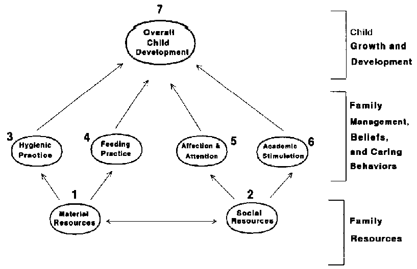
It is a well known fact that the family is pivotal in determining the general wellbeing of an individual (United Nations University, 2011). Patients with EDS are no exception to the effect of the family on their health condition. Here, I will consider the structural model of family and social health/child development so as to understand how the family affects the wellbeing of an EDS’ patient (United Nations University, 2011.

As it can be seen in the model above, material and social resources are primary in determining the overall well being of a child. For an EDS’ patient, material resources are prerequisite for medical costs and professional advice. As it is normally the case, the quality of medical care that can be provided to a patient will always be proportional to available material resources. Since EDS is a condition that requires the extension of medical care to the home, material resources are prerequisite here. The provision of high hygiene standards and prescribed diets to a patient is obviously important for the management of EDS. Since mental and emotional health is almost synonymous with physical health, social resources (from affected family) are crucial in enhancing the management of EDS. Provision of affection, among other emotional needs, is prerequisite for enhancing the wellbeing of someone with EDS (United Nations University, 2011).
Epidemiology in the Community
As I had stated earlier, the prevalence of EDS has been estimated at about 1: 25000 (Wenstrup, 2007). The above a ratio applies in the United States, as well as in other parts of the world (Callewaert, 2008). Currently, no research has been done to establish the prevalence of EDS in specific ethnic communities (Callewaert, 2008). It is important to note that the prevalence of EDS could be significantly higher than stated above; since there could be a high number of cases that have gone unreported (Callewaert, 2008). Among the various types of EDS, the most serious type that often leads to premature deaths is the vascular type. Vascular EDS has a prevalence of about 5-10% globally (Wenstrup, 2007).
Conclusion
Ehlers danlos syndrome is an inheritable disease that manifests itself in various forms. Considering the various complications that can arise due to EDS, it is important for suspect patients to seek prerequisite medical attention for management. An appropriate medical package would include a differential process to determine a specific EDS in a patient, an implementation of appropriate laboratory tests if necessary, and importantly, a management and treatment program.
Reference List
Callewaert, B. (2008). Ehlers-Danlos syndromes and marfan syndrome. Best Practice & Research Clinical Rheumatology 22, 1,165-189
Fransiska, M. (2010) Clinical and genetic aspects of Ehlers-Danlos syndrome Genetics in Medicine, 12 (10), 597-600.
Germain, P. D. (2007). Ehlers Danlos syndrome-type IV Orphanet Journal of Rare Diseases 2 (32), 152-210.
Groft, S. (2010). Rare diseases epidemiology Advances in Experimental Medicine 686, 354-68
United Nations University. (2011). Structural models of family social health theory. Web.
Watanabe, A. (2008). The Vascular type of Ehlers Danlos Syndrome J Nippon Medical School 75 (5), 125-180.
Wenstrup, R. (2007). Ehlers danlos syndrome: classical type. Seattle: University of Washington.