Abstract
Ultrasound is a widely used imaging technique in medicine either as a therapeutic or as a diagnostic tool. Ultrasound imaging technique is used in almost every form of medicine ranging from cardiology, internal medicine, anesthesia, dentistry, hypothermia, physical therapy, surgery etc. Millions of ultrasound scans are done every year, and ultrasound remains one of the fastest growing imaging techniques in medicine. The popularity of ultrasound imaging in medicine is due to the facts that it is a low cost, real time and less risky technique. The wide usage of ultrasound in medical imaging elicits the question of the biological effects of ultrasound examination on living tissues.
This paper evaluates the basic physics behind the use of ultrasound imaging technique with an emphasis on the medical ultrasound imaging. The paper also evaluates the physical mechanisms for the biological effects of ultrasound and the effects of ultrasound on living tissues in vivo and vitriol. Several effects of ultrasound on tissues are then reviewed to provide an insight on the range of the effects of ultrasound on tissues.
This report also explores the significance of the biological effects of ultrasound on the safe use of ultrasound techniques in medical practice. Concerning the exposure of humans to ultrasound, the paper covers the applications of ultrasound on the human medicine. This report is intended to cover the biological effects of ultrasound with peculiar interest in clinical applications. This report, therefore, be considered a valuable resource for those interested in the use of ultrasound imaging and its effects.
Introduction
Ultrasound involves the use of sound waves of high intensity to create images of body organs and the body systems. An ultra sound machine produces various images that allow the organs and systems in the body to be visualized. The ultrasound machine sends out sound waves of high frequency that reflect off the body structures. A computer then receives the waves reflected and uses them to create a picture of the body tissues. There is no ionizing radiation used in ultrasound imaging. The use of ultrasound in medical diagnosis is called ultrasonography (NCRP, 2006).
An ultrasound technician can use information from an ultrasound image to answer questions about a medical condition. A transducer also called prove is used to project and then receive the reflected sound waves. A gel is normally applied on the patient’s skin so as not to distort the waves as they are crossing the skin. Ultrasound scans can answer questions about a medical condition using an understanding of human anatomy and the picture of the reflected waves (Andreassi, 2004).
The widespread use of ultrasound imaging in medicine and diagnostics raised concerns over the effects of the use of this technique on the body. Concerns have been raised in the past over whether there are any adverse effects of the sound waves in the body. Experiments carried out on animals have provided a link to ultrasound in cancers, internal bleeding, schizophrenia and autism (Nelson & Pretorus, 2008).
However, although there are some biological effects of ultrasound on tissues, no adverse affects have been reported to date over the use of ultrasound in medical imaging. Some of the biological effects of ultrasound on tissues are transient and not well understood. It is also difficult to perform valid experimental procedures on the effects of ultrasound on tissues and, therefore, the use of ultrasound technique has been limited to the use of as low as reasonably active (ALARA) principle (Guy van, Steven, & Cozy, 2007).
Physics background of ultrasound
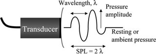
Sound is a form of mechanical energy and propagates longitudinally in elastic media in alternating zones of rarefaction and compression. Ultrasound imaging uses short pulses of sound, and the energy is reflected in impulses. The basic properties of sound waves used in medical imaging are of frequency 1 to 15 MHz, pulse length of 3 to five cycles, the wavelength is 05mm, and attenuation is one db the speed of sound in tissue is 1540m/s. The pulses reflected in moving interfaces like blood vessels exhibit a shift in phase that can be used to measure the velocity of the movement along the sound beam. These are called the Doppler Effect and are in the range of 10 to 1000 Hz (Barry, 2011).
There are two types of Doppler. The velocity Doppler computes the velocity of each pixel and displays color schemes whose direction and saturation depends on the component measured. The power Doppler computes the entire velocity distribution and displays the magnitude as either color or brightness (Dalecki, 2004).

Ultrasound also uses contrast materials such as gas filled micro bubbles for better visualization of tissues and vessels. These contrast agents help in enhancing the masses, visualizing blood flow and delivering drugs and genetic agents to sites. The sound beam used in ultrasound propagates in two directions, the Fresnel field and the far flied. The scan lines sweeping in different directions are used to create a dimensional image of the tissue under analysis (Collins, 2008). The potential of an ultrasound beam to cause bio effects is indicated by the formula:
- ISPPA=ISATA/duty cycle
An Ipa=ita/duty cycle, is the indicator of the potential, thermal, effect of ultrasound.
- Ispta=Isata (Isp/Isa) duty cycle
This is the indicator of the potential mechanical bio effects and the cavitations of ultrasound.
The potential bio effects of ultrasound are also influenced by the power output and the acoustic power of the ultrasound machine i.e. the rate of production of energy, flow and absorption. Different tissues of the body have different attenuation coefficients for sound waves. For example, the lung has an attenuation coefficient of 40, while the blood has an attenuation coefficient of 0.18. This different attenuation coefficient of tissues determines the frequency and the intensity of ultrasound scan that is used in different tissues of the body (Collins, 2008).
Mechanism of interaction of ultrasound with body tissues and the potential bio effects
Ultrasound is a form of mechanical energy where pressurized sound waves travel through tissues. Reflected and scattered waves are then used to form an image of the object under scan (Nelson & Pretorius, 2008). The major effects, of ultrasound are characterized into:
Thermal effects
As the ultrasound sound waves travel through tissue, their energy is converted into heat in tissues. Tissues with high absorption coefficient like bone have highest absorption and record high thermal effects than tissues with low absorption coefficients like amniotic fluids. The Energy conversion of sound waves in tissues is also dependent on tissue thermal characteristics, the intensity of ultrasound and the period of exposure. The intensity of ultrasound is in turn dependent on power output, the mode of ultrasound, the depth of scanning and area of imaging. There are so many variables involved in thermal effects of sound waves on tissues that it is difficult to model temperature rises in tissue. The transducer phase of ultrasound can also heat in an ultrasound examination (NCRP, 2006).
Cavitations
Cavitations’ is the development of stable or temporal gas bubbles in tissues. Inertial (transient cavitations have the most damage on tissue because when the gas filled cavity grows, then during pressure rarefaction of the ultrasound pulse and contraction during the compression phase the collapse of the bubble can generate high pressure and high temperatures. This has been hypothesized to be the cause of hemorrhage in lungs and intestines of experimental animals. The use of contrast agents in ultrasound leads to the formation of micro bubbles that in a way provide nuclei for cavitations to form (Dalecki, 2004).
Other mechanical effects
Passage of ultrasound waves on tissues causes a low form of radiation force on tissue. However, the pressure that result from ultrasound waves and the pressure gradient that develops across the beam of sound even at high intensities of the diagnostic range are exceptionally low. The effect of this radiation force manifest itself best in fluid volumes where streaming can develop within the fluid. However, the fluid velocities that may develop are unlikely to cause any damage in tissues (Nyborg, 2011).
The effect of ultrasound on fetus
No evidence of cavitations on fetal scanning but hyperthermia has been thought to be tetranogenic. However, the results are not particularly clear and valid to indicate any adverse effect of ultrasound on fetus. The guidelines for use of ultrasound in pregnancy are that ultrasound scans that results in temperature rise, in tissue of 1.5degres centigrade, may be used without reservations and those above 4 degrees centigrade are potentially hazardous to tissue (Colins, 2008).
Chemical effects
Experimental ultrasound can lead to depolymerisation of DNA, polysaccharide and polypeptides. Oxidation and reduction reactions can also occur although these processes have not been reported in vivo, in diagnostic procedures. High intensity and high frequency ultrasound have been shown to result in chromosomal; damage, genetic mutations and tissue necrosis and teratogenic effects (Barry, 2011).
Applications in medicine and biology
Ultrasound has been used in many clinical settings for medical imaging. The main advantage of ultrasound over other diagnosis and therapeutic procedures is that certain structures of the body can be visualized without using radiation. Ultrasound is also faster than x-rays or other radiographic techniques (NCRP, 2006).
The Use of ultra sound in diagnosis
Obstetrics
Ultrasound imaging is routinely used in examining the progression of pregnancy, and observation of tumors in ovary, uterus and fallopian tubes. Ultrasound is the most common procedure used in measuring the size of the fetus, the position of the fetus in the uterus and checking the position of the placenta to assess whether it is developing properly over the opening of the cervix (Nelson & Pretorius, 2008).
In cardiology, ultrasound imaging is used to examine the heart function and blood flow motion in a procedure called echocardiography. Ultrasound is also used to assess the functioning of many body organs in orthopedics and blood vessel diseases (Barry, 2011). Ultrasound is also used in therapeutics. It may help physicians insert needles into the body. In urology ultrasound, imaging is used in measuring blood flow in kidneys, visualization of kidney stones and detection of prostrate cancer. Ultrasound is also finding uses as a diagnostic tool in emergency rooms (Dalecki, 2004).
Summary and recommendations
The use of ultrasound in diagnosis and therapeutics has provided a wealth of knowledge in medicine, and there is a need to appreciate the impact that the technique has had on medicine. Millions of ultrasound examinations are done every year, and ultra sonography is one of the fastest growing imaging techniques. The growth of ultrasound technique in imaging is partly, because of this it is a low cost real time image display, and largely the few bio effects it has on body tissues.
Although animal studies have shown that the ultrasound procedure has some potential bio effects on tissue, the regulatory processes that control the use of ultrasound devices has set a safety margin for the safe use of ultrasound in clinical settings since high intensity high frequency sound waves are damaging to the body. These guidelines have restricted the patient exposure to ultrasound procedures to be restricted to levels that produce little or no bio effects (Nyborg, 2011). The regulations stipulating the use of ultrasound devices are set by bodies like the International Electro Technical Association, the USA food and drugs administration and the federation of societies of ultrasound in Medicine and biology (EFSUMB) (Nyborg, 2011).
The field of ultrasound imaging is rapidly changing. Therefore, a physician or an Ultrasonographer is needed to play a leading role in adhering to the accepted exposure limits of ultrasound to limit the potential effects of ultrasound on tissues. This calls for prudent adherence to the standards of safe use of ultrasound by the physician and the ultrasound technicians.
References
Andreassi, M. (2004). The biological effects of diagnostic cardiac imaging on chronically Exposed Physicians. The importance of being none ionizing. Pisa: Institute of Clinical Physiology.
Barry, G. (2011). Ultrasound bio effects for the perinatologist. Web.
Collin, D. (2008). Safety of diagnostic ultrasound in fetal scanning. London: Centrus.
Dalecki, D. (2004). Mechanical bioEffects of ultrasound. Annual review of biomedical Engineering. Volume 6, 229-248.
George, L. (2011). The physics of ultrasound. Sydney: BAT Research group.
Guy van, C., Steven, D., Cosyn, B. (2007). Bio effects of ultrasound contrast Agents in Daily Clinical practice: fact or fiction. European heart journal. vol. 102, 224-254.
Nelson, T., & Pretorius, D. (2008). Ultrasound bio effects. Web.
NCRP, (2006). Biological effects of ultrasound mechanisms and clinical implications. Milan: NCRP.
Nyborg, W. (2011). The biological effects of ultrasound: development of safety Guidelines. Part ii General Review. Burlington: University of Vermont.